A Case of Anterograde Amnesia with Bilateral Hippocampus Involvement After Acute Glufosinate Ammonium Intoxication
- Affiliations
-
- 1Department of Radiology, Catholic University of Daegu Medical School, Daegu, Korea. ysw.badest.2010@gmail.com
- 2Department of Radiology, Kyungpook National University Medical School, Daegu, Korea.
- KMID: 2206932
- DOI: http://doi.org/10.13104/jksmrm.2014.18.4.352
Abstract
- A 51-year-old man developed anterograde amnesia following the ingestion of glufosinate ammonium. Brain MRI revealed hyperintense lesions involving the bilateral hippocampus and parahippocampal gyrus, and the right occipital lobe. The mechanism underlying acute glufosinate ammonium intoxication and the differential diagnosis of hippocampal lesions are discussed.
Keyword
MeSH Terms
Figure
-
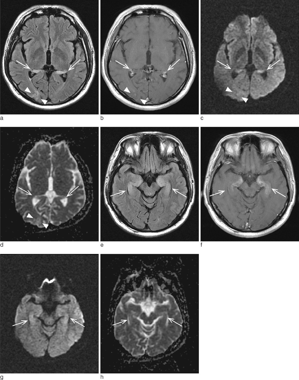
Fig. 1 Brain MRI of a 51-year-old man on the 10th day following glufosinate ammonium intoxication (a-d, basal ganglia level; e-h, midbrain level). (a) Fluidattenuated inversion recovery (FLAIR) image and (b) gadolinium-enhanced T1-weighted imaging (Gd-T1WI) at the basal ganglia level showing bilateral hyperintensities and parenchymal enhancement in the bilateral hippocampus (arrows) and right occipital lobe (arrowheads). This lesion exhibited hyperintensities on (c) diffusion-weighted imaging (DWI) and (d) apparent diffusion coefficient (ADC) mapping, suggesting vasogenic edema. The same pattern was observed in the bilateral hippocampus and parahippocampal gyrus (arrows) on (e) FLAIR, (f) Gd-T1WI, (g) DWI, and (h) ADC mapping at the midbrain level.
Cited by 1 articles
-
Anterograde Amnesia after Acute Glufosinate Ammonium Intoxication
Hyuk-Hoon Kim, Young-Gi Min
Acute Crit Care. 2018;33(2):110-113. doi: 10.4266/acc.2016.00444.
Reference
-
1. Watanabe T, Sano T. Neurological effects of glufosinate poisoning with a brief review. Hum Exp Toxicol. 1998; 17:35–39.2. Park HY, Lee PH, Shin DH, Kim GW. Anterograde amnesia with hippocampal lesions following glufosinate intoxication. Neurology. 2006; 67:914–915.3. Lee HY, Song SY, Lee SH, Lee SY, Kim SH, Ryu SW. Vasogenic edema in striatum following ingestion of glufosinate-containing herbicide. J Clin Neurosci. 2009; 16:1372–1373.4. Förster A, Griebe M, Gass A, Kern R, Hennerici MG, Szabo K. Diffusion-weighted imaging for the differential diagnosis of disorders affecting the hippocampus. Cerebrovasc Dis. 2012; 33:104–115.5. Cha SY, Kang TH, Kim SJ, et al. Selective anterograde amnesia associated with hippocampal and splenial damage after heat stroke. Clin Neurol Neurosurg. 2013; 115:1867–1870.6. Park JS, Kwak SJ, Gil HW, Kim SY, Hong SY. Glufosinate herbicide intoxication causing unconsciousness, convulsion, and 6th cranial nerve palsy. J Korean Med Sci. 2013; 28:1687–1689.7. Kim TY, Park DW, Park CK, Lee YJ, Lee SR. Reversible splenial lesion in the corpus callosum on MRI after ingestion of a herbicide containing glufosinate ammonium: a case report. J Korean Soc Radiol. 2014; 70:399–402.8. Nakaki T, Mishima A, Suzuki E, Shintani F, Fujii T. Glufosinate ammonium stimulates nitric oxide production through N-methyl D-aspartate receptors in rat cerebellum. Neurosci Lett. 2000; 290:209–212.9. Matsumura N, Takeuchi C, Hishikawa K, Fujii T, Nakaki T. Glufosinate ammonium induces convulsion through N-methyl-D-aspartate receptors in mice. Neurosci Lett. 2001; 304:123–125.10. Waxman EA, Lynch DR. N-methyl-D-aspartate receptor subtypes: multiple roles in excitotoxicity and neurological disease. Neuroscientist. 2005; 11:37–49.11. Calas AG, Richard O, Même S, et al. Chronic exposure to glufosinate-ammonium induces spatial memory impairments, hippocampal MRI modifications and glutamine synthetase activation in mice. Neurotoxicology. 2008; 29:740–774.12. Meme S, Calas AG, Montécot C, et al. MRI characterization of structural mouse brain changes in response to chronic exposure to the glufosinate ammonium herbicide. Toxicol Sci. 2009; 111:321–330.
- Full Text Links
-
- Actions
-
Cited
- CITED
-
- Close
- Share
-
- Similar articles
-
- Anterograde Amnesia after Acute Glufosinate Ammonium Intoxication
- Diffusion-Weighted Imaging for Detecting Glufosinate Ammonium Intoxication: A Case Report
- Encephalopathy After Glufosinate Ammonium Intoxication
- Erythema Multiforme-like Eruptions Caused by Accidental Exposure to Glufosinate Ammonium
- Two Cases of Acute Glufosinate Ammonium Intoxication with Disparate Outcomes
