Brain MRI Findings of Welders: High Signal Intensity in T1W1 Secondary to Manganese Exposure
- Affiliations
-
- 1Department of Diagnostic Radiology, Sunlin Presbyterian Hospital.
- 2Department of Occupational medicine, Sunlin Presbyterian Hospital.
- 3Department of Neurology, Sunlin Presbyterian Hospital.
- 4Department of Preventive medicine, Medical College, Dongguk University.
- KMID: 2201393
- DOI: http://doi.org/10.3348/jkrs.1998.38.3.403
Abstract
-
PURPOSE: To evaluate the clinical and brain MRI findings of welders and to determine the utility of MRI inthe assessment of occupational manganese exposure
MATERIALS AND METHODS
Eighteen welders, working non-regularly,and three of their wives were examined. Clinical, neurologic and brain MRI findings were retrospectively analyzed.Blood manganese(Mn), urine Mn, serum iron(Fe) and blood cadmium (Cd) were measured. The mean period of exposurewas 16(7-32) years. High signal intensity of the globus pallidus(GP). as seem on T1-Weighted images(T1WI), wasgraded as follows : Grade 1, when its signal intensity (SI) was slighty higher than that of frontal subcorticalwhite matter; Grade 3, when the SI of GP was as high as that of subcutaneous fat ; and grade 2 for intermediatecases. We calculated the pallidal index (SI of GP/SI of frontal subcortical white matter) and correlated this withblood and urine Mn levels.
RESULTS
All welders complained of fatigue, headache, anorexia, and decreased libido.The palmomental reflex was positive in five(28%), Myerson's sign in four(22%), and intention tremor in three(17%).Mean blood Mn was 5.18(range, 1.77-9.34)microgram/dl, mean urine Mn was 5.84(range, 1.07-22/microgram/l, serum Fe was elevatedin one welder, and serum Cd in two. T1WI of brain MRI revealed high signal intensities in the globus pallidus, theputamen, the substantia nigra, the tectum, the caudate nucleus, the subthalamic nucleus, the hypothlamus along theextrapyramidal tract, and the pituitary gland. T2WI of brain MRI revealed no significant signal change. PI and GPgrading showed good correlation(kappa=0.60). Blood Mn correlated closely with the pallidal index(Rs=0.48, P=0.046),but there was no correlation between urine Mn and the pallidal index(Rs=0.1>0.05).
CONCLUSION
T1WI of brain MRIin welders showed high signal intensities in the globus pallidus, the putamen, the tectum, the caudate nucleus,the subthalamic nucleus, the hypothalamus and the pituitary gland. These intensities correlated closely with bloodMn levels, suggesting their potential role in estimating the accumulation of Mn in the brain.
MeSH Terms
Figure
-
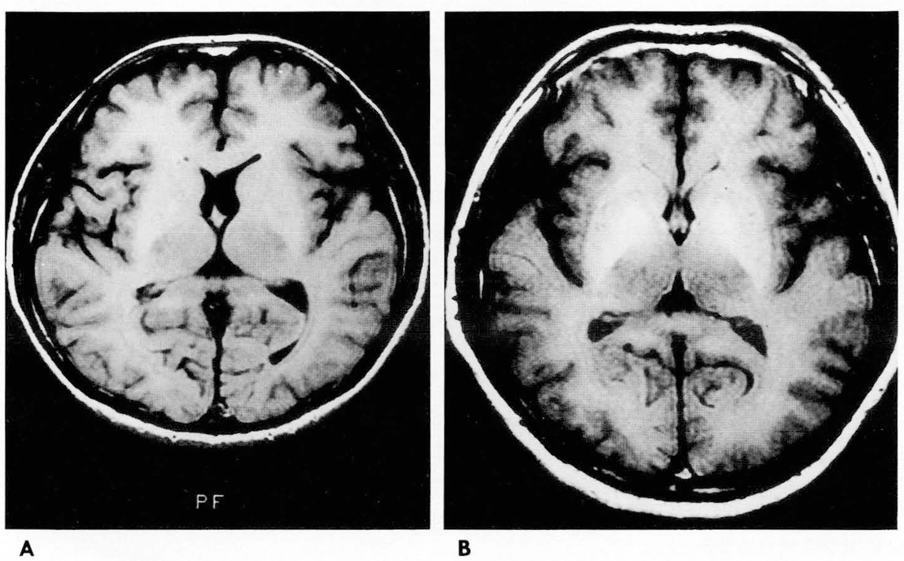
Fig. 1. Grading of high signal intensity of the globus pallidus on Tl- weighted MR image A. Axial Tl-weighted MR image (T1WI) (600/25) shows signal intensity of globus pallidus slightly higher than that of the frontal subcortical white matter (grade 1) B. Axial T1WI (600/25) shows high signal intensity of the globus pallidus similar to that of the subcutaneous fat(grade 3).

Fig. 2. Brain MRI of a welder with 12 years’ exposure to manganese with headache and fatigue A. Axial T1WI (600/25! shows symmetrical hyperintensities in the globus pallidus(grade 3) and the pu- tamen(grade 2). The caudate nucleus and the thalamus exhibit normal signal intensity. B. Axial T1WI (600/25) shows symmetrical hyperintensities in bilateral substantia nigra(grade 2). C. Sagittal T1WI (600/25) shows hyperintensity of the entire pituitary gland(grade 3), the hypothala- mus(grade 2), the subthalamic nu- cleus(grade 2), and the midbrain tectum(grade 2). D. Axial T2WI (2300/90) shows no evidence of significant signal change in any areas.

Fig. 3. Disappearance of high signal intensity after cessation of welder's work. Initial axial TlWIs (A, B) (600/20) in a welder with 10 years’ exposure history shows symmetrical grade 2 hyperintensity in the globus pallidus and grade 1 hyperintensity in the putamen(A), and grade 2 hyperintensity in the substantia nigra(B). Follow-up axial TlWIs (C, D) (600/ 20) obtained 7 months after withdrawal from exposure to manganese without medical treatment reveal near complete disappearance of high signal intensities in the globus pallidus, the putamen(C), and the midbrain(D). Blood level of manganese also decreased.
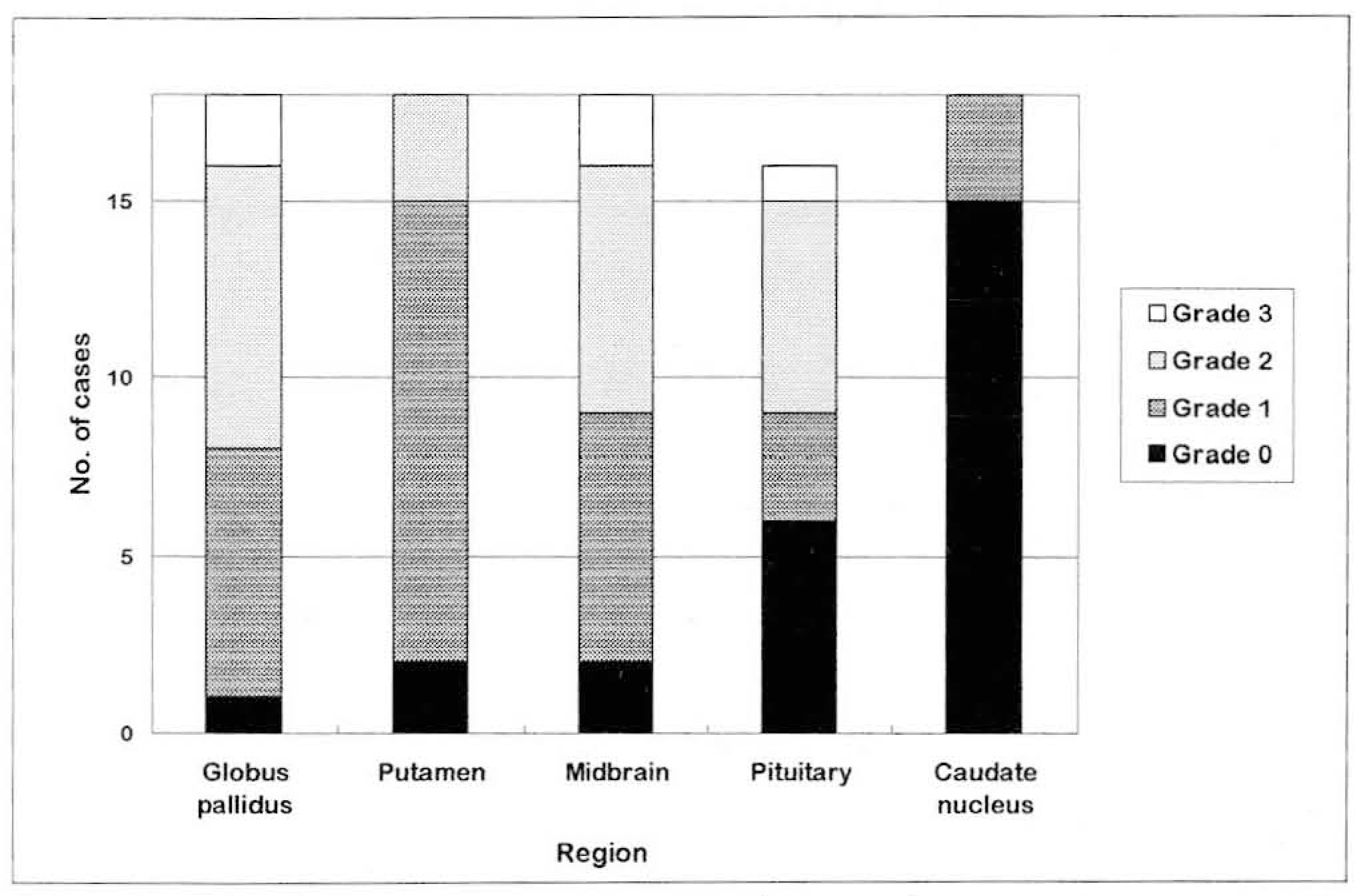
Fig. 4. Number of cases by grading of signal intensity in each region.
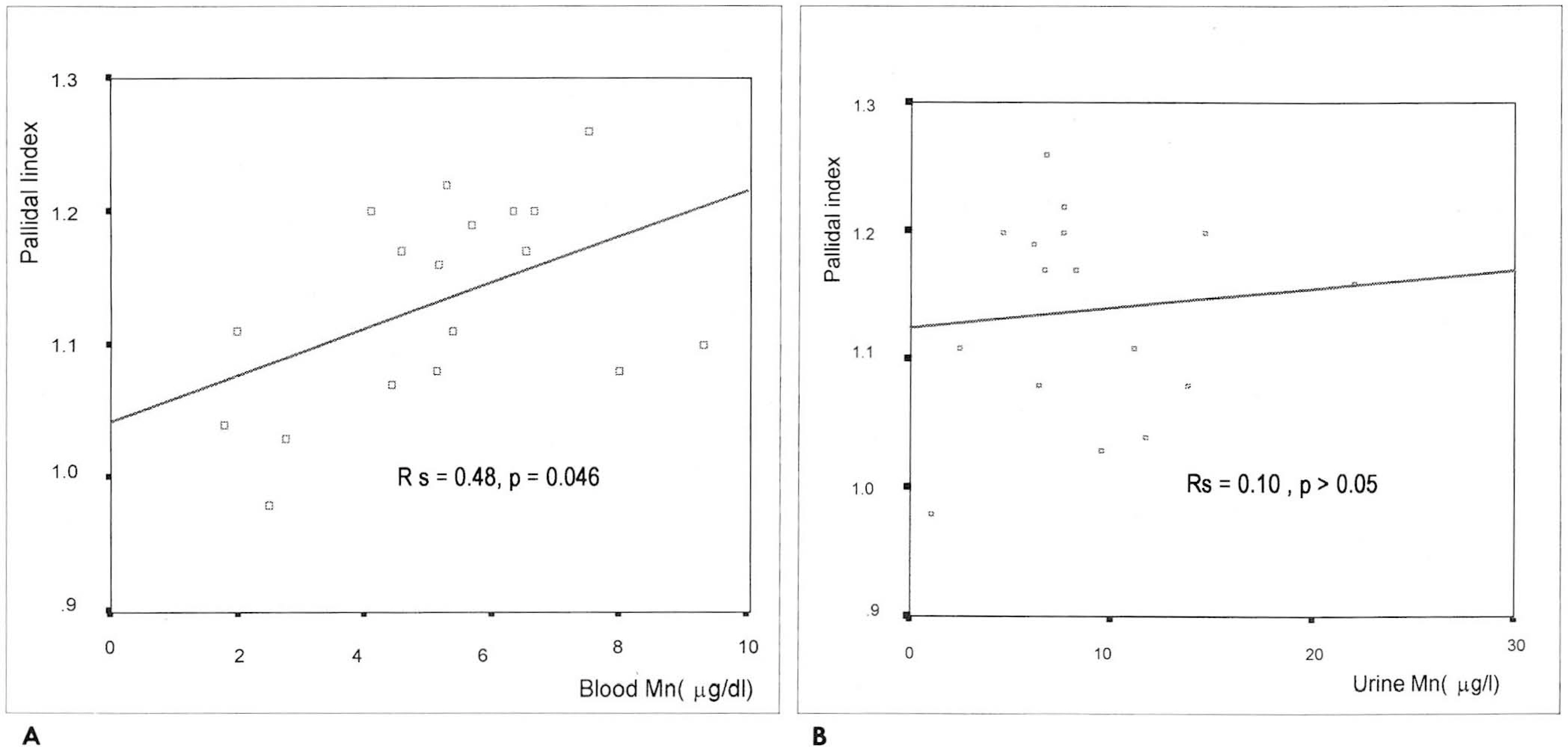
Fig. 5. Correlation between pallidal index and blood and urine manganese levels. A. Pallidal index is significantly correlated with blood manganese level(Spearmann rank correlation coefficient of 0.48) (Rs=0.48, P=0.046). B. Pallidal index does not show significant correlation with urinary manganese level(Spearmann rank correlation coefficient of 0.10)(Rs=0.10, Ρ) 0.05).
Reference
-
1.Guidotti TL. Hazards of welding technologies. In Rom WN. Environmental and occupational medicine. 2nd ed.Little, Brown and Company;1992. p. 831–841.2.Sjogren B. Effects of gases and particals in welding and soldering. In Zenz C, Dickerson OB, eds. Occupational medicine. 3rd ed.Mosby;1994. p. 917–924.3.Shinotoh H., Snow BJ., Hewitt KA, et al. MRI and PET studies of manganese-intoxicated monkeys. Neurology. 1995. 45:1199–1204.
Article4.Nelson K., Golnick J., Korn T., Angle C. Manganese encephalopathy: Utility of early magnetic resonance imaging. Br J Ind Med. 1993. 50:510–513.
Article5.Calne DB., Chu NS., Huang CC., Lu CS., Olanow W. Manganism and idiopathic parkinsonism: Similarities and differences. Neurology. 1994. 44:1583–1586.
Article6.Mirowitz SA., Westrich TJ. Basal ganglial signal intensity alterations: Reversal after discontinuation of parenteral manganese administration. Radiology. 1992. 185:535–536.
Article7.Gianutsos G., Seltzer MD., Saymeh R., Wu MW., Michel RG. Brain manganese accumulation following systemic accumulation of different forms. Arch Toxicol. 1985. 57:272–275.8.Scheuhammer AM., Cherian MG. The influence of manganese on the distribution of manganese, magnesium, zinc, iron, and copper in rats after chronic manganese exposure. J Toxicol Environ Health. 1983. 12:361–370.9.Krieger D., Krieger S., Jansen O, et al. Manganese and chronic hepatic encephalopathy. Lancet. 1995. 346:270–274.
Article10.Inoue E., Hori S., Narumi Y, et al. Portal-systemic encephalopathy: Presence of basal ganglia lesions with high signal intensity on MR Images. Radiology. 1991. 179:551–555.
Article11.Pujol A., Pujol J., Graus F, et al. Hyperintense globus pallidus on Τ1-weighted MRI in cirrhotic patients is associated with severity of liver failure. Neurology. 1993. 43:65–69.12.Mirowitz SA., Westrich TJ., Hirsch JD. Hyperintense basal ganglia on Τ1-weighted MR Images in patients receiving parenteral nutrition. Radiology. 1991. 181:117–120.13.Montgomery EB. Heavy metals and the etiology of parkinson's disease and other movement disorders. Toxicology. 1995. 97:3–9.
Article14.Newland MC., Ceckler TL., Kordower JH., Weiss B. Visualizing manganese in the primate basal ganglia with magnetic resonance imaging. Exp Neurol. 1989. 106:251–258.
Article15.백승국, 안우현, 최한용, 김봉기. T1 강조영상에서뇌기저핵의고신호강도. 대한방사선의학회지. 1994. 30(1):1–5.16.김윤주, 최순정, 김창수, 김선희, 정순필, 김양숙. 뇌기저핵부위의고신호강도: 간경변환자에서간문맥전신성뇌증과의연관성. 대한방사선의학희지. 1993. 29(1):33–37.17.Henkelman RM., Watts JF., Kucharczyk W. High signal intensity in MR images of calcified brain tissue. Radiology. 1991. 179:199–206.
Article18.Mirowitz SA., Sartor K., Gado M. High signal basal ganglia lesions on Τ1-weighted images in neurofibromatosis. AJR. 1990. 154:369–373.19.Barkovich AJ. MR and CT evaluation of profound systemic and infantile asphyxia. AJNR. 1992. 13:959–972.20.Osborn AG. Inherited metabolic, white matter, and degenerative disease of the brain. In Osborn AG eds. Diagnostic neuroradiology. 1st ed.Mosby;1994. p. 717–747.21.Cuesta MC., Gomez G., Bonilla E., Suarez-Roca H. Autoreceptor presynaptic control of dopamine release from striatum is lost at early stages of manganese poisoning. Life Science. 1995. 56:1857–1864.22.Graham. D. G. Catecholamine toxicity: a proposal for the molecular pathogenesis of manganese neurotoxicity and parkinson's disease. Neurotoxicology. 1984. 5:83–96.23.Agil A., Fuller CJ., Jialal I. Susceptibility of plasma to ferrous iron/hydrogen peroxide-mediated oxidation: demonstration of a possible Fenton reaction. Clin Chem. 1995. 41:220–225.
Article24.Presti D., Scott JE. Hyaluronan-mediated protective effect against cell damage caused by enzymatically produced hydroxyl (OH')radicals is dependent on hyaluronan molecular mass. Cell Biochem Fund. 1994. 15:2475–2478.25.Lexa FJ., Trojanowski JQ., Braffman BH., Atlas SW. The aging brain and neurodegenerative disease. In Atlas SW, ed. Magnetic resonance image of brain and spine. Philadelphia & NewYork: Lippincott-Raven. 1996. 842–852.
- Full Text Links
-
- Actions
-
Cited
- CITED
-
- Close
- Share
-
- Similar articles
-
- Study on Clinical Significance of High Signal Intensity by Brain Magnetic Resonance Imaging in Mild Steel/Arc Welders (Clinical Significance of High Signal Intensity by Brain MRI in Welders)
- An Association between Brain MRI and Neurologic Findings in Welders Exposed to Manganese Fume
- Significance of Brain Magnetic Resonance Imaging (MRI) in the Assessment of Occupational Manganese Exposure
- A study on manganese health hazards among experienced welders
- The Significance of Increased Signal Intensity in MR Imaging among Male Welders
