Comparison of occusal aspects in monolithic zirconia crown before and after occlusal adjustment during intraoral try-in: a case report
- Affiliations
-
- 1Department of Prosthodontics, School of Dentistry, Yonsei University, Seoul, Republic of Korea. jfshim@yuhs.ac
- KMID: 2195438
- DOI: http://doi.org/10.4047/jkap.2014.52.3.246
Abstract
- In case of prosthesis fabrication by CAD/CAM, location, area and contour of occlusal contacts can be adjusted so more functional occlusion can be acquired. Also, errors in a manufacturing process is reduced compared to cast metal prostheses and porcelain fused metal prostheses fabricated by conventional methods such as casting and porcelain build up. Therefore, prostheses by CAD/CAM show superior occlusion accuracy. Recently, virtual articulator function has been introduced to CAD/CAM system, which reproduces mandibular movement against maxilla. Thus, it is possible to consider occlusal interference in anterior/lateral movement as well as closing movement. There have been many studies on the marginal and internal fit of prostheses using zirconia but the occlusal fit of zirconia crown fabricated by CAD/CAM has not been researched as much. In this case report, 7 zirconia crowns were designed and fabricated by CAD/CAM for total 5 patients. The models of zirconia crowns before and after occlusal adjustment during intraoral try-in were scanned for occlusal contacts, which were compared to evaluate accuracy of prostheses and understand patterns of occlusal adjustment. Most of the occlusal adjustments were done on functional cusps and slopes of zirconia crown, and the magnitude of occlusal adjustment ranged from 15 microm to 60 microm. In the zirconia crown fabricated with CAD/CAM systems, the occlusal adjustment is a necessary procedure, so additional procedures will be needed for compensating reduced mechanical properties.
Keyword
MeSH Terms
Figure
-

Fig. 1. The overlapped image between CAD designed image and model scanned image of left lower 1 st molar. (A) The original overlapped data. (B) The color map for indicating the changed amount of occlusal surface. ∗m = mesial, d = distal, b = buccal, l = lingual.
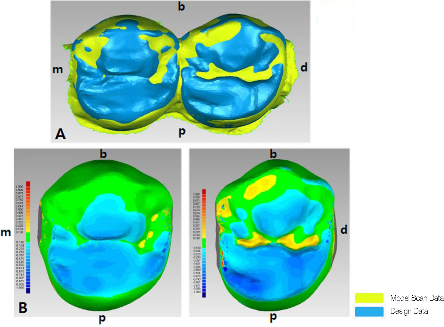
Fig. 2. The overlapped image between CAD designed image and model scanned image of left upper 1 st premolar and 2 nd premolar. (A) The original overlapped data. (B) The color map for indicating the changed amount of occlusal surface. ∗m = mesial, d = distal, b = buccal, p = palatal.
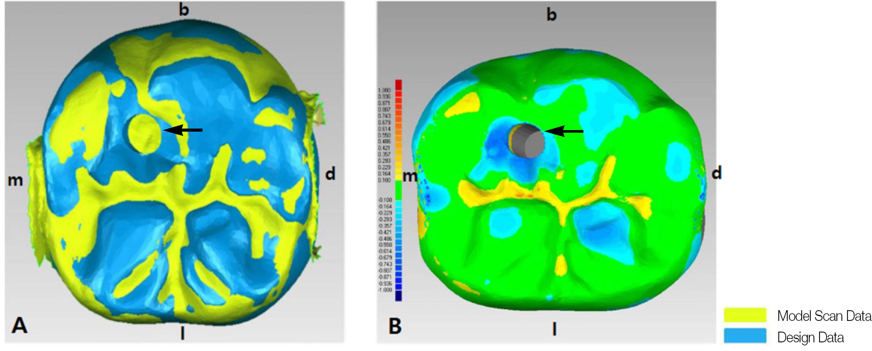
Fig. 3. The overlapped image between CAD designed image and model scanned image of right lower 1 st molar. (A) The original overlapped data. (B) The color map for indicating the changed amount of occlusal surface. ∗m = mesial, d = distal, b = buccal, l = lingual, arrow = implant screw hole.
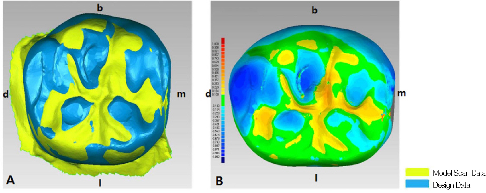
Fig. 4. The overlapped image between CAD designed image and model scanned image of left lower 1 st molar. (A) The original overlapped data. (B) The color map for indicating the changed amount of occlusal surface. ∗m = mesial, d = distal, b = buccal, l = lingual.

Fig. 5. The overlapped image between CAD designed image and model scanned image of right lower 1 st molar and 2 nd molar. (A) The original overlapped data. (B) The color map for indicating the changed amount of occlusal surface. ∗m = mesial, d = distal, b = buccal, l = lingual.
Reference
-
1. Fukui K, Kaneuji A, Sugimori T, Ichiseki T, Kitamura K, Matsumoto T. Wear comparison between a highly cross-linked polyethylene and conventional polyethylene against a zirconia femoral head: minimum 5-year follow-up. J Arthroplasty. 2011; 26:45–9.2. Oilo M, Kvam K, Gjerdet NR. Simulation of clinical fractures for three different all-ceramic crowns. Eur J Oral Sci. 2014; 122:245–50.
Article3. Maestre-Ferr l′ n L, Romero-Milla′n J, Pen ̃arrocha-Oltra D, Pen ̃arrocha-Diago M. Virtual articulator for the analysis of dental occlusion: an update. Med Oral Patol Oral Cir Bucal. 2012; 17:e160–3.4. Scotti R, Cardelli P, Baldissara P, Monaco C. Clinical fitting of CAD/CAM zirconia single crowns generated from digital intraoral impressions based on active wavefront sampling. J Dent. 2011 Oct 17.
Article5. Kohorst P, Junghanns J, Dittmer MP, Borchers L, Stiesch M. Different CAD/CAM-processing routes for zirconia restorations: influence on fitting accuracy. Clin Oral Investig. 2011; 15:527–36.
Article6. Lughi V, Sergo V. Low temperature degradation-aging- of zirconia: A critical review of the relevant aspects in dentistry. Dent Mater. 2010; 26:807–20.7. Preis V, Behr M, Hahnel S, Handel G, Rosentritt M. In vitro failure and fracture resistance of veneered and full-contour zirconia restorations. J Dent. 2012; 40:921–8.
Article
- Full Text Links
-
- Actions
-
Cited
- CITED
-
- Close
- Share
-
- Similar articles
-
- Superimposition: a simple method to minimize occlusal adjustment of monolithic restoration
- Comparison of interference from eccentric movements of dental crowns fabricated via dynamic jaw motion tracking and conventional methods: a double-blind clinical study
- Fracture strength of zirconia monolithic crowns
- Maxillary cement retained implant supported monolithic zirconia prosthesis in a full mouth rehabilitation: a clinical report
- Fracture strength of zirconia monolithic crowns and metal-ceramic crowns after cyclic loading and thermocycling
