J Korean Neurosurg Soc.
2015 Jun;57(6):469-472. 10.3340/jkns.2015.57.6.469.
Intracranial Undifferentiated Sarcoma Arising from a Low-Grade Glioma: A Case Report and Literature Review
- Affiliations
-
- 1Department of Neurosurgery, Korea University Guro Hospital, Seoul, Korea. ns806@kumc.or.kr
- KMID: 2191257
- DOI: http://doi.org/10.3340/jkns.2015.57.6.469
Abstract
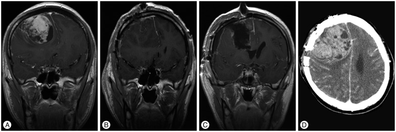
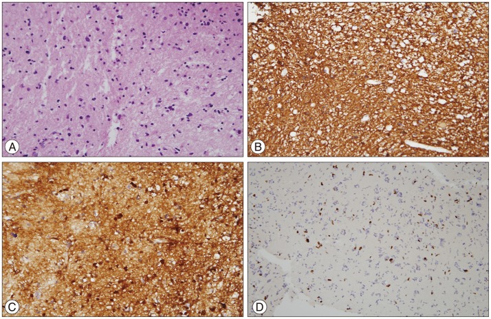
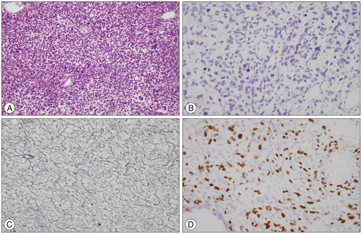
- Undifferentiated sarcomas are rarely identified in the intracranial region. A 23-year-old man was admitted with a chief complaint of headache. Initial magnetic resonance images showed signs of low-grade glioma in the frontal lobe. Stereotactic biopsy was performed, and a diagnosis of diffuse astrocytoma was confirmed. Three months later, the patient presented with a high-grade tumor as seen on imaging studies. He underwent total resection of the tumor and histopathological tests identified an undifferentiated sarcoma. The patient died eight months later due to massive tumor bleeding. To the best of our knowledge, this is the first report of undifferentiated sarcoma arising from low-grade glioma without any chemotherapy or radiotherapy.
Keyword
MeSH Terms
Figure
Reference
-
1. Alexandru D, Linskey ME, Kim RC, Bota D. Undifferentiated sarcoma induced by BCNU (1,3-bis(2-chloroethyl)-1-nitrosourea) wafers. J Interdiscipl Histopathol. 2013; 1:163–167.
Article2. Brennan MF, Antonescu CR, Maki RG. Undifferentiated Pleomorphic Sarcoma (UPS; Malignant Fibrous Histiocytoma : MFH) and Myxofibrosarcoma. In : Brennan MF, Antonescu CR, Maki RG, editors. Management of Soft Tissue Sarcoma. ed 1. New York: Springer Science & Business Media;2013. p. 129–136.3. Fletcher CD. The evolving classification of soft tissue tumours - an update based on the new 2013 WHO classification. Histopathology. 2014; 64:2–11. PMID: 24164390.
Article4. Fletcher CD. Undifferentiated sarcomas : what to do? And does it matter? A surgical pathology perspective. Ultrastruct Pathol. 2008; 32:31–36. PMID: 18446665.
Article5. Hofer SO, Shrayer D, Reichner JS, Hoekstra HJ, Wanebo HJ. Wound-induced tumor progression : a probable role in recurrence after tumor resection. Arch Surg. 1998; 133:383–389. PMID: 9565118.6. Kosucu P, Yavuz AA, Yavuz MN, Reis A, Ahmetoglu A. Primary intracranial malignant fibrous histiocytoma : case report. Eur J Radiol Extra. 2003; 47:78–81.7. Lehnhardt M, Daigeler A, Homann HH, Schwaiberger V, Goertz O, Kuhnen C, et al. MFH revisited : outcome after surgical treatment of undifferentiated pleomorphic or not otherwise specified (NOS) sarcomas of the extremities-an analysis of 140 patients. Langenbecks Arch Surg. 2009; 394:313–320. PMID: 18584203.
Article8. Meng X, Zhong J, Liu S, Murray M, Gonzalez-Angulo AM. A new hypothesis for the cancer mechanism. Cancer Metastasis Rev. 2012; 31:247–268. PMID: 22179983.
Article9. Mitsuhashi T, Watanabe M, Ohara Y, Hatashita S, Ueno H. Multifocal primary intracerebral malignant fibrous histiocytoma-case report. Neurol Med Chir (Tokyo). 2004; 44:249–254. PMID: 15200060.
Article10. Nascimento AF, Raut CP. Diagnosis and management of pleomorphic sarcomas (so-called "MFH") in adults. J Surg Oncol. 2008; 97:330–339. PMID: 18286476.
Article11. Sasagawa Y, Tachibana O, Iizuka H. Undifferentiated sarcoma of the cavernous sinus after γ knife radiosurgery for pituitary adenoma. J Clin Neurosci. 2013; 20:1152–1154. PMID: 23587603.
Article12. Schmitt WR, Carlson ML, Giannini C, Driscoll CL, Link MJ. Radiation-induced sarcoma in a large vestibular schwannoma following stereotactic radiosurgery : case report. Neurosurgery. 2011; 68:E840–E846. discussion E846PMID: 21209595.13. Weiss SW, Enzinger FM. Malignant fibrous histiocytoma : an analysis of 200 cases. Cancer. 1978; 41:2250–2266. PMID: 207408.
Article14. Yang T, Rockhill J, Born DE, Sekhar LN. A case of high-grade undifferentiated sarcoma after surgical resection and stereotactic radiosurgery of a vestibular schwannoma. Skull Base. 2010; 20:179–183. PMID: 21318035.
Article15. Yoo RE, Choi SH, Park SH, Jung HW, Kim JH, Sohn CH, et al. Primary intracerebral malignant fibrous histiocytoma : CT, MRI, and PET-CT findings. J Neuroimaging. 2013; 23:141–144. PMID: 21447025.
Article16. Yun LG, Shin WH, Kim BT, Choi SK, Byun BJ, Lee DW. Malignant fibrous histiocytoma presenting as osteolytic lesion on the left temporal bone : a case report. J Korean Neurosurg Soc. 1995; 24:471–476.
- Full Text Links
- Actions
-
Cited
- CITED
-
- Close
- Share
- Similar articles
-
- Low-Grade Fibromyxoid Sarcoma Arising in Posterior Nasal Cavity: Case Report and Review of the Literature
- Two Cases of Low Grade Endometrial Stromal Sarcoma
- Chordoid Glioma: an Uncommon Tumor of the Third Ventricle
- A Case of Recurrent Low Grade Endometrial Stromal Sarcoma in the Ureter
- A case of primary retroperitoneal undifferentiated endometrial stromal sarcoma after concurrent chemoradiation therapy for cervical cancer


