J Korean Neurosurg Soc.
2014 Jun;55(6):331-336. 10.3340/jkns.2014.55.6.331.
Comparison of Sagittal Spinopelvic Alignment between Lumbar Degenerative Spondylolisthesis and Degenerative Spinal Stenosis
- Affiliations
-
- 1Department of Neurosurgery, Kyung Hee University Hospital at Gangdong, Kyung Hee University School of Medicine, Seoul, Korea. spinekim@khu.ac.kr
- KMID: 2191112
- DOI: http://doi.org/10.3340/jkns.2014.55.6.331
Abstract
OBJECTIVE
The purpose of this study was to evaluate the differences in sagittal spinopelvic alignment between lumbar degenerative spondylolisthesis (DSPL) and degenerative spinal stenosis (DSS).
METHODS
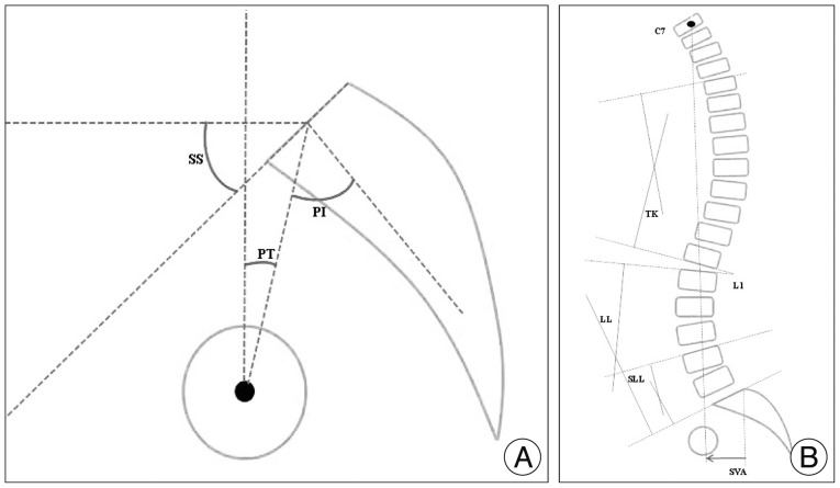
Seventy patients with DSPL and 72 patients with DSS who were treated with lumbar interbody fusion surgery were included in this study. The following spinopelvic parameters were measured on whole spine lateral radiographs in a standing position : pelvic incidence (PI), pelvic tilt (PT), sacral slope (SS), lumbar lordosis angle (LL), L4-S1 segmental lumbar angle (SLL), thoracic kyphosis (TK), and sagittal vertical axis from the C7 plumb line (SVA). Two groups were subdivided by SVA value, respectively. Normal SVA subgroup and positive SVA subgroup were divided as SVA value (<50 mm and > or =50 mm). Spinopelvic parameters/PI ratios were assessed and compared between the groups.
RESULTS
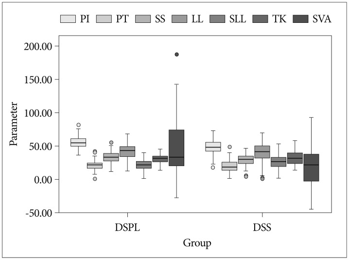
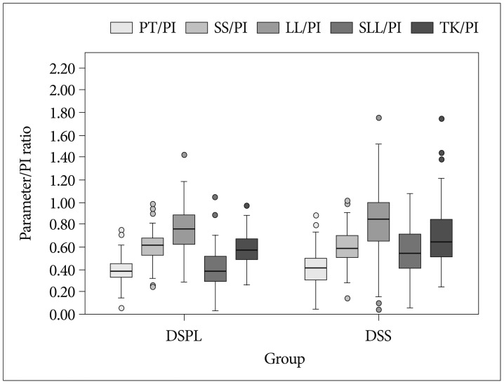
The PI of DSPL was significantly greater than that of DSS (p=0.000). The SVA of DSPL was significantly greater than that of DSS (p=0.001). In sub-group analysis between the positive (34.3%) and normal SVA (65.7%), there were significant differences in LL/PI and SLL/PI (p<0.05) in the DSPL group. In sub-group analysis between the positive (12.5%) and normal SVA (87.5%), there were significant differences in PT/PI, SS/PI, LL/PI and SLL/PI ratios (p<0.05) in the DSS group.
CONCLUSION
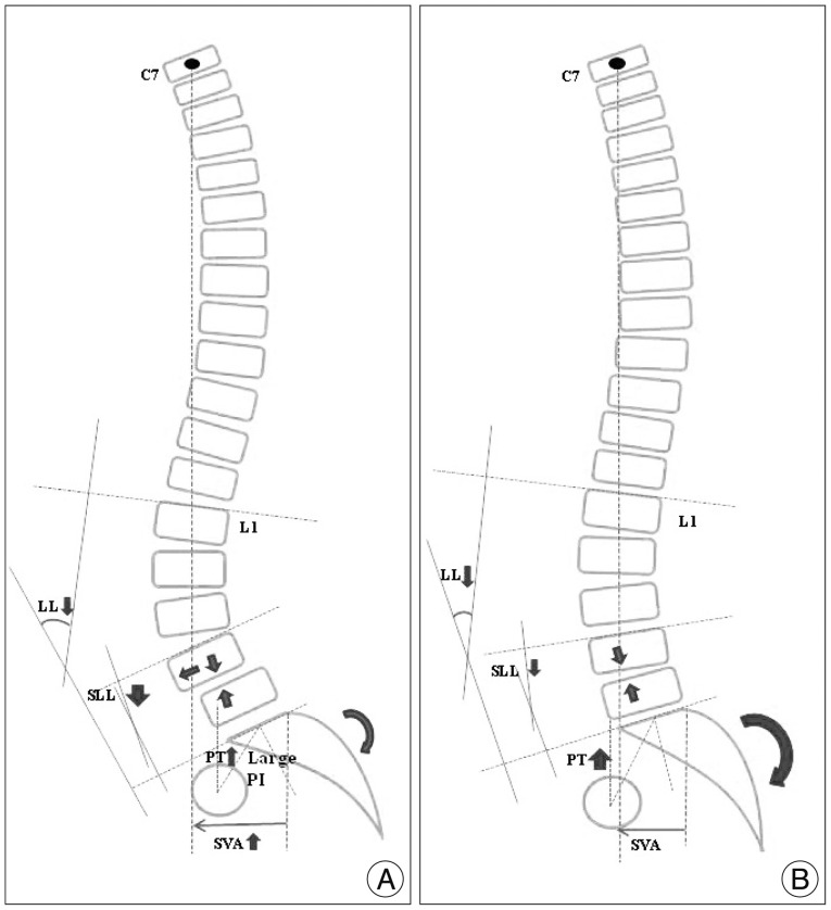
Patients with lumbar degenerative spondylolisthesis have the propensity for sagittal imbalance and higher pelvic incidence compared with those with degenerative spinal stenosis. Sagittal imbalance in patients with DSPL is significantly correlated with the loss of lumbar lordosis, especially loss of segmental lumbar lordosis.
Keyword
MeSH Terms
Figure
Reference
-
1. Arnoldi CC, Brodsky AE, Cauchoix J, Crock HV, Dommisse GF, Edgar MA, et al. Lumbar spinal stenosis and nerve root entrapment syndromes. Definition and classification. Clin Orthop Relat Res. 1976; (115):4–5. PMID: 1253495.2. Barrey C, Jund J, Noseda O, Roussouly P. Sagittal balance of the pelvis-spine complex and lumbar degenerative diseases. A comparative study about 85 cases. Eur Spine J. 2007; 16:1459–1467. PMID: 17211522.
Article3. Barrey C, Jund J, Perrin G, Roussouly P. Spinopelvic alignment of patients with degenerative spondylolisthesis. Neurosurgery. 2007; 61:981–986. discussion 986. PMID: 18091275.
Article4. Curylo LJ, Edwards C, DeWald RW. Radiographic markers in spondyloptosis : implications for spondylolisthesis progression. Spine (Phila Pa 1976). 2002; 27:2021–2025. PMID: 12634562.5. Duval-Beaupère G, Schmidt C, Cosson P. A Barycentremetric study of the sagittal shape of spine and pelvis : the conditions required for an economic standing position. Ann Biomed Eng. 1992; 20:451–462. PMID: 1510296.
Article6. Funao H, Tsuji T, Hosogane N, Watanabe K, Ishii K, Nakamura M, et al. Comparative study of spinopelvic sagittal alignment between patients with and without degenerative spondylolisthesis. Eur Spine J. 2012; 21:2181–2187. PMID: 22639298.
Article7. Hammerberg EM, Wood KB. Sagittal profile of the elderly. J Spinal Disord Tech. 2003; 16:44–50. PMID: 12571484.
Article8. Hanson DS, Bridwell KH, Rhee JM, Lenke LG. Correlation of pelvic incidence with low- and high-grade isthmic spondylolisthesis. Spine (Phila Pa 1976). 2002; 27:2026–2029. PMID: 12634563.
Article9. Jackson RP, Kanemura T, Kawakami N, Hales C. Lumbopelvic lordosis and pelvic balance on repeated standing lateral radiographs of adult volunteers and untreated patients with constant low back pain. Spine (Phila Pa 1976). 2000; 25:575–586. PMID: 10749634.
Article10. Jackson RP, McManus AC. Radiographic analysis of sagittal plane alignment and balance in standing volunteers and patients with low back pain matched for age, sex, and size. A prospective controlled clinical study. Spine (Phila Pa 1976). 1994; 19:1611–1618. PMID: 7939998.
Article11. Korovessis P, Dimas A, Iliopoulos P, Lambiris E. Correlative analysis of lateral vertebral radiographic variables and medical outcomes study short-form health survey : a comparative study in asymptomatic volunteers versus patients with low back pain. J Spinal Disord Tech. 2002; 15:384–390. PMID: 12394662.
Article12. Labelle H, Roussouly P, Berthonnaud E, Transfeldt E, O'Brien M, Chopin D, et al. Spondylolisthesis, pelvic incidence, and spinopelvic balance : a correlation study. Spine (Phila Pa 1976). 2004; 29:2049–2054. PMID: 15371707.13. Legaye J, Duval-Beaupère G, Hecquet J, Marty C. Pelvic incidence : a fundamental pelvic parameter for three-dimensional regulation of spinal sagittal curves. Eur Spine J. 1998; 7:99–103. PMID: 9629932.
Article14. Lim JK, Kim SM. Difference of sagittal spinopelvic alignments between degenerative spondylolisthesis and isthmic spondylolisthesis. J Korean Neurosurg Soc. 2013; 53:96–101. PMID: 23560173.
Article15. Meyerding H. Spondylolisthesis. Surg Gynecol Obstet. 1932; 54:371–377.16. Oh SK, Chung SS, Lee CS. Correlation of pelvic parameters with isthmic spondylolisthesis. Asian Spine J. 2009; 3:21–26. PMID: 20404942.
Article17. Pearson A, Blood E, Lurie J, Tosteson T, Abdu WA, Hillibrand A, et al. Degenerative spondylolisthesis versus spinal stenosis : Degenerative spondylolisthesis versus spinal stenosis : does a slip matter? Comparison of baseline characteristics and outcomes (SPORT). Spine (Phila Pa 1976). 2010; 35:298–305. PMID: 20075768.18. Rosenberg NJ. Degenerative spondylolisthesis. Predisposing factors. J Bone Joint Surg Am. 1975; 57:467–474. PMID: 1141255.
Article19. Schuller S, Charles YP, Steib JP. Sagittal spinopelvic alignment and body mass index in patients with degenerative spondylolisthesis. Eur Spine J. 2011; 20:713–719. PMID: 21116661.
Article20. Vaz G, Roussouly P, Berthonnaud E, Dimnet J. Sagittal morphology and equilibrium of pelvis and spine. Eur Spine J. 2002; 11:80–87. PMID: 11931071.
Article
- Full Text Links
- Actions
-
Cited
- CITED
-
- Close
- Share
- Similar articles
-
- MR Findings of Spondylolisthesis: Assessment of Associated Spinal and Neural Foraminal Stenosis
- Degenerative Spondylolisthesis in Thoracic Spine
- Difference of Sagittal Spinopelvic Alignments between Degenerative Spondylolisthesis and Isthmic Spondylolisthesis
- The Relationship between Sagittal Spinal Alignment and Surgical Results in Degenerative Lumbar Scoliosis with Spinal Stenosis
- A Clinical Study on the Causes of the Nerve Entrapment in the Degenerative Spondylolisthesis


