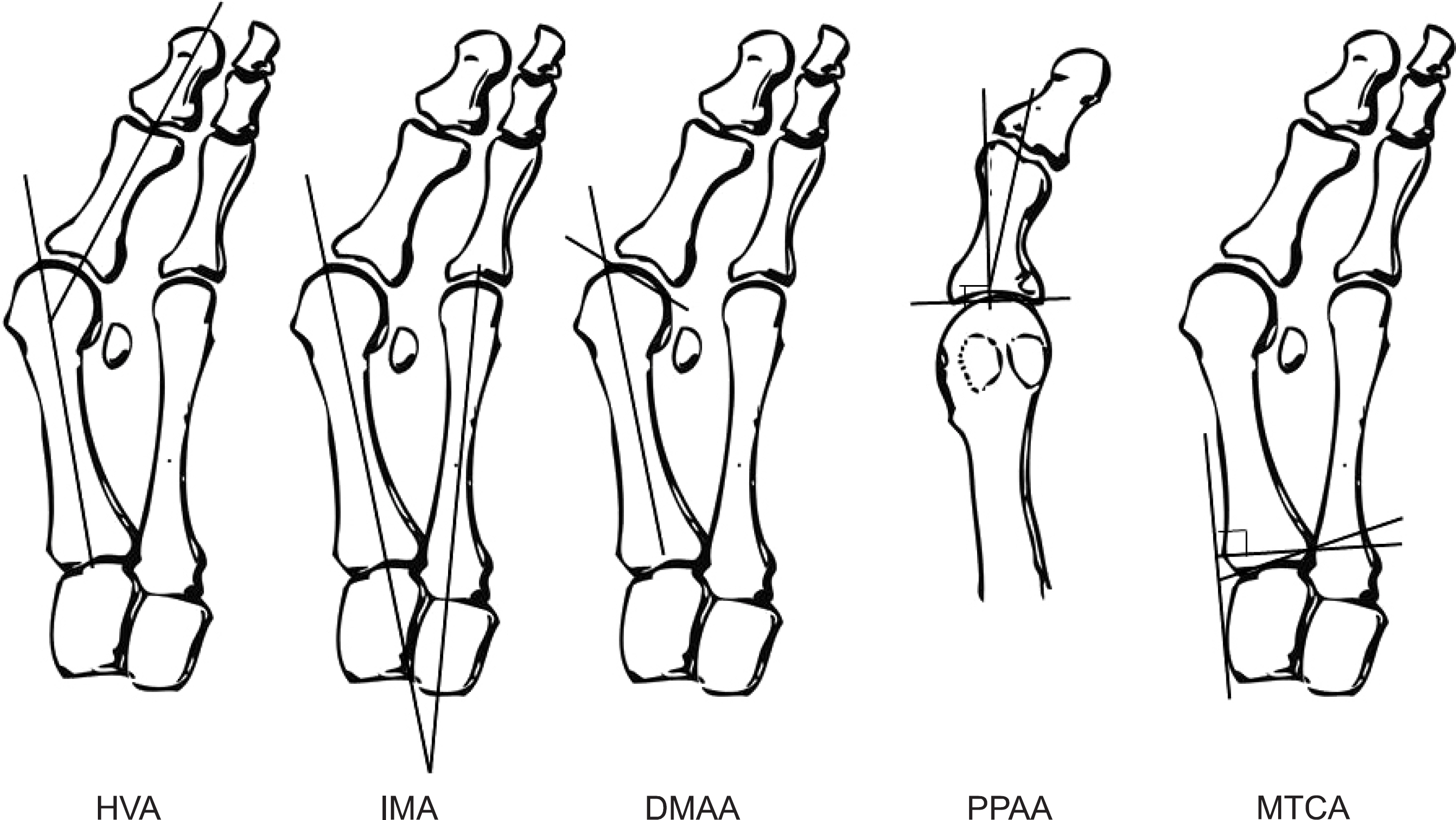
J Korean Foot Ankle Soc.
2014 Jun;18(2):43-47. 10.14193/jkfas.2014.18.2.43.
Diagnosis and Pathophysiology of Hallux Valgus
- Affiliations
-
- 1Department of Orthopedic Surgery, Korea University Guro Hospital, Korea University College of Medicine, Seoul, Korea. dakjul@hanmail.net
- KMID: 2181659
- DOI: http://doi.org/10.14193/jkfas.2014.18.2.43
Abstract
- Hallux valgus is a lateral deviation of the first phalanx and medial deviation of the first metatarsal at the first metatarsophalangeal (MP) joint. Its incidence has increased due to developing footwear. The etiologies include fashion footwear, genetic causes, anatomical abnormality around the foot, rheumatoid arthritis, and neuromuscular disorders. Physiologic alignment of the first MP joint is maintained by congruent and symmetric alignment of the articular surface of the first proximal phalanx and first metatarsal head, physiologic relationship of the distal first metatarsal articular surface and the first metatarsal shaft axis, and stable balance of soft tissue around the first MP joint and stable tarsometatarsal joint. Several factors have been associated with hallux valgus, including pes planus, hypermobility of the first tarsometatarsal joint, flattened shape of the first metatarsal head, increased distal metatarsal articular angle, and deformation of the medial capsular integrity. History and physical examination are very important to diagnosis of hallux valgus. Simple radiography provides information on deformity, particularly in weight-bearing anteroposterior and lateral radiographs. Understanding the etiologies and pathophysiology is very important for success in treatment of patients with hallux valgus.
Keyword
MeSH Terms
Figure
Cited by 2 articles
-
Approach for the Treatment on Hallux Valgus
Sung-Hyun Lee, Yeong-Chang Lee
J Korean Foot Ankle Soc. 2019;23(4):143-148. doi: 10.14193/jkfas.2019.23.4.143.Current Trends in the Treatment of Hallux Valgus: Analysis of the Korean Foot and Ankle Society (KFAS) Member Survey
Jaeho Cho, Byung-Ki Cho, Hyun-Woo Park, Ki-Sun Sung, Su-Young Bae
J Korean Foot Ankle Soc. 2021;25(4):157-164. doi: 10.14193/jkfas.2021.25.4.157.
Reference
-
References
1. Mann RA, Coughlin MJ. Adult hallux valgus. Coughlin MJ, Mann RA, editors. Surgery of the foot and ankle. 7th ed.St. Louis: Mosby Inc.;1999. p. 150–269.2. Coughlin MJ, Jones CP. Hallux valgus: demographics, etiology, and radiographic assessment. Foot Ankle Int. 2007; 28:759–77.
Article3. Coughlin MJ, Thompson FM. The high price of high-fashion footwear. Instr Course Lect. 1995; 44:371–7.4. Scott G, Wilson DW, Bentley G. Roentgenographic assessment in hallux valgus. Clin Orthop Relat Res. 1991; 267:143–7.
Article5. Yoo CI, Kim BH, Shin KS, Im JI. A clinical and radiological study of the hallux valgus angle, intermetatarsal angle and hallux valgus of Koreans. J Korean Orthop Assoc. 1990; 25:1183–90.6. Easley ME, Trnka HJ. Current concepts review: hallux valgus part 1: pathomechanics, clinical assessment, and nonoperative management. Foot Ankle Int. 2007; 28:654–9.
Article7. Kato T, Watanabe S. The etiology of hallux valgus in Japan. Clin Orthop Relat Res. 1981; 157:78–81.
Article8. Sim-Fook L, Hodgson AR. A comparison of foot forms among the non-shoe and shoe-wearing Chinese population. J Bone Joint Surg Am. 1958; 40:1058–62.
Article9. Mann RA, Coughlin MJ. Hallux valgus–etiology, anatomy, treatment and surgical considerations. Clin Orthop Relat Res. 1981; 157:31–41.
Article10. Craigmile DA. Incidence, origin, and prevention of certain foot defects. Br Med J. 1953; 2:749–52.
Article11. Kalen V, Brecher A. Relationship between adolescent bunions and flatfeet. Foot Ankle. 1988; 8:331–6.
Article12. Kilmartin TE, Wallace WA. The significance of pes planus in juvenile hallux valgus. Foot Ankle. 1992; 13:53–6.
Article13. Ledoux WR, Shofer JB, Smith DG, Sullivan K, Hayes SG, Assal M, et al. Relationship between foot type, foot deformity, and ulcer occurrence in the high-risk diabetic foot. J Rehabil Res Dev. 2005; 42:665–72.
Article14. Saragas NP, Becker PJ. Comparative radiographic analysis of parameters in feet with and without hallux valgus. Foot Ankle Int. 1995; 16:139–43.
Article15. Faber FW, Kleinrensink GJ, Verhoog MW, Vijn AH, Snijders CJ, Mulder PG, et al. Mobility of the first tarsometatarsal joint in relation to hallux valgus deformity: anatomical and biomechanical aspects. Foot Ankle Int. 1999; 20:651–6.
Article16. Shi K, Hayashida K, Tomita T, Tanabe M, Ochi T. Surgical treatment of hallux valgus deformity in rheumatoid arthritis: clinical and radiographic evaluation of modified Lapidus technique. J Foot Ankle Surg. 2000; 39:376–82.
Article17. Myerson MS, Badekas A. Hypermobility of the first ray. Foot Ankle Clin. 2000; 5:469–84.18. Faber FW, Kleinrensink GJ, Mulder PG, Verhaar JA. Mobility of the first tarsometatarsal joint in hallux valgus patients: a radiographic analysis. Foot Ankle Int. 2001; 22:965–9.
Article19. Coughlin MJ, Shurnas PS. Hallux valgus in men. Part II: first ray mobility after bunionectomy and factors associated with hallux valgus deformity. Foot Ankle Int. 2003; 24:73–8.
Article20. Doty JF, Coughlin MJ. Hallux valgus and hypermobility of the first ray: facts and fiction. Int Orthop. 2013; 37:1655–60.
Article21. Schneider W. Influence of different anatomical structures on distal soft tissue procedure in hallux valgus surgery. Foot Ankle Int. 2012; 33:991–6.
Article22. Young KW, Kim JS, Cho JW, Lee KW, Park YU, Lee KT. Characteristics of male adolescent-onset hallux valgus. Foot Ankle Int. 2013; 34:1111–6.
Article23. Nery C, Coughlin MJ, Baumfeld D, Ballerini FJ, Kobata S. Hallux valgus in males–part 2: radiographic assessment of surgical treatment. Foot Ankle Int. 2013; 34:636–44.24. Uchiyama E, Kitaoka HB, Luo ZP, Grande JP, Kura H, An KN. Pathomechanics of hallux valgus: biomechanical and immunohistochemical study. Foot Ankle Int. 2005; 26:732–8.
Article25. Fuhrmann RA, Layher F, Wetzel WD. Radiographic changes in forefoot geometry with weightbearing. Foot Ankle Int. 2003; 24:326–31.
Article26. Tanaka Y, Takakura Y, Takaoka T, Akiyama K, Fujii T, Tamai S. Radiographic analysis of hallux valgus in women on weightbearing and nonweightbearing. Clin Orthop Relat Res. 1997; 336:186–94.
Article
- Full Text Links
- Actions
-
Cited
- CITED
-
- Close
- Share
- Similar articles
-
- Corrective Osteotomies in Hallux Valgus
- Incidence of Hallux Valgus Interphalangeus in the Normal and Hallux Valgus Feet and its Correlations with Hallux Valgus Angle and Intermetatarsal Angle
- Consideration of Various Medial Capsulorrhaphy Methods in Hallux Valgus Surgery
- Technique Tip: A Simple Method to Treat Hallux Valgus with Severe Metatarsus Adductus
- Arthrodesis Of The First Metsatarophalangeal Joint For Rheumatoid Arthritis And Hallux Valgus


