Full mouth rehabilitation with a few remaining teeth and implants for a patient with chronic periodontitis: a case report
- Affiliations
-
- 1Department of Prosthodontics, School of Dentistry, Chonnam National University, Gwangju, Republic of Korea. yhsdent@jnu.ac.kr
- 2RIS Foundation for Advanced Biomaterials, School of Dentistry, Chonnam National University, Gwangju, Republic of Korea.
- KMID: 2180027
- DOI: http://doi.org/10.14368/jdras.2015.31.3.253
Abstract
- Chronic periodontitis involves subsequent loss of teeth, and if left untreated, can lead to adjacent teeth drifting and supraeruption of the rest dentition. Careful consideration has to be given when deciding extraction of remaining teeth in treatment of periodontally compromised dentitions. For tooth-supported fixed partial dentures or removable partial dentures, periodontally compromised teeth are extracted due to possible early failure from functional overload, but for implant restoration, the teeth could be used as supports for fixed partial dentures because implants can reduce overload on teeth. The remaining natural teeth can help clinicians restoring vertical dimension and normal occlusal plane in full mouth rehabilitation because it conserves patients' proprioceptive response. This clinical report describes treatment of a patient who has a few remaining teeth and supraeruption of the rest dentition from severe chronic periodontitis. Satisfactory clinical result was achieved with full mouth rehabilitation using a few teeth and implants.
MeSH Terms
Figure
-

Fig. 1 Panogramic radiograph at first visit; Multiple teeth loss, shifting teeth, supraeruption of the rest dentition and severe periodontitis.
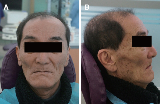
Fig. 2 Extraoral photos at first visit. (A) Frontal view, (B) Side view.

Fig. 3 Implant installation; Panoramic view. #17i, 16i, 45i, 47i: US II Φ5.0 × 11 mm, #14i, 24i: US II Φ4.0 × 10 mm, #26i, 27i: US II Φ4.5 × 11 mm, #33i, 43i: US II Φ3.5 × 11 mm, 13i, 11i, 23i: TS Φ3.5 × 11.5 mm (Osstem, Busan, Korea).
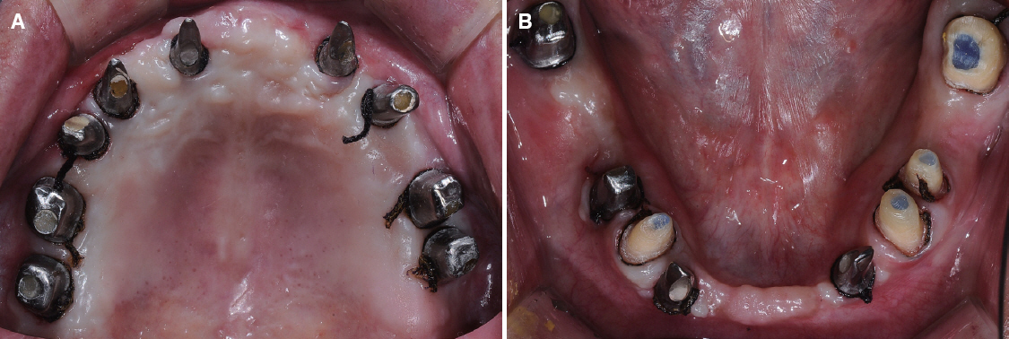
Fig. 4 Impression for provisional restoration. (A) Maxillary occlusal view, (B) Mandibular occlusal view.

Fig. 5 Provisional restoration. (A) Balancing side during left lateral excursion, (B) Maxillary occlusal view, (C) Working side during left lateral excursion, (D) Right buccal view during centric occlusion, (E) Frontal view during centric occlusion, (F) Left buccal view during centric occlusion, (G) Working side during right lateral excursion, (H) Mandibular occlusal view, (I) Balancing side during right lateral excursion.
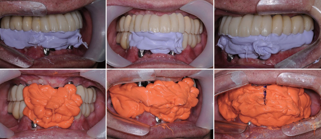
Fig. 6 Intermaxillary relation registration for cross mounting.
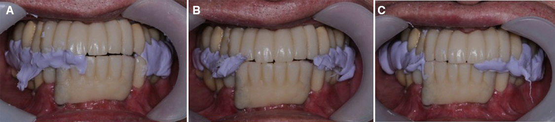
Fig. 7 Check bite registration. (A) Left excursion, (B) Anterior guidance, (C) Right excursion.

Fig. 8 Mounting on a semi-adjustable dental articulator. (A) Facebow transfer, (B) Cross mounting on a articulator.

Fig. 9 Definitive restoration. (A) Balancing side during left lateral excursion, (B) Maxillary occlusal view, (C) Working side during left lateral excursion, (D) Right buccal view during centric occlusion, (E) Frontal view during centric occlusion, (F) Left buccal view during centric occlusion, (G) Working side during right lateral excursion, (H) Mandibular occlusal view, (I) Balancing side during right lateral excursion.

Fig. 10 CT for condylar joint space. (A) Right condyle, (B) Left condyle.
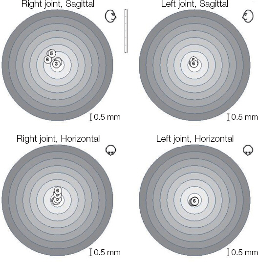
Fig. 11 EPA test by ARCUS digma II. MICP: 1, 2, 3; CR (manipulation): 4, 5, 6.

Fig. 12 Postoperative panoramic radiograph.

Fig. 13 Postoperative extraoral photos. (A) Frontal view, (B) Side view.
Reference
-
References
1. Lundgren D, Nyman S, Heijl L, Carlsson GE. Functional analysis of fixed bridges on abutment teeth with reduced periodontal support. J Oral Rehabil. 1975; 2:105–16. DOI: 10.1111/j.1365-2842.1975.tb01521.x. PMID: 1095705.2. Nyman S, Lindhe J. A longitudinal study of combined periodontal and prosthetic treatment of patients with advanced periodontal disease. J Periodontol. 1979; 50:163–9. DOI: 10.1902/jop.1979.50.4.163. PMID: 374703.3. Mengel R, Behle M, Flores-de-Jacoby L. Osseointegrated implants in subjects treated for generalized aggressive periodontitis: 10-year results of a prospective, long-term cohort study. J Periodontol. 2007; 78:2229–37. DOI: 10.1902/jop.2007.070201. PMID: 18052693.4. Hoffmann O, Beaumont C, Zafiropoulos GG. Combined periodontal and implant treatment of a case of aggressive periodontitis. J Oral Implantol. 2007; 33:288–92. DOI: 10.1563/1548-1336(2007)33[288:CPAITO]2.0.CO;2.5. Ekelund JA, Lindquist LW, Carlssson GE, Jemt T. Implant treatment in the edentulous mandible: a prospective study on Brånemark system implants over more than 20 years. Int J Prosthodont. 2003; 16:602–8. PMID: 14714838.6. Attard NJ, Zarb GA. Long-term treatment outcomes in edentulous patients with implant-fixed prostheses: the Toronto study. Int J Prosthodont. 2004; 17:417–24. PMID: 15382777.7. Berglundh T, Persson L, Klinge B. A systematic review of the incidence of biological and technical complications in implant dentistry reported in prospective longitudinal studies of at least 5 years. J Clin Periodontol. 2002; 29(Suppl 3):197–212.8. Fransson C, Lekholm U, Jemt T, Berglundh T. Prevalence of subjects with progressive bone loss at implants. Clin Oral Implants Res. 2005; 16:440–6. DOI: 10.1111/j.1600-0501.2005.01137.x. PMID: 16117768.9. Roos-Jansåker AM, Lindahl C, Renvert H, Renvert S. Nine- to fourteen-year follow-up of implant treatment. Part II: presence of peri-implant lesions. J Clin Periodontol. 2006; 33:290–5. DOI: 10.1111/j.1600-051X.2006.00906.x. PMID: 16553638.10. Schou S. Implant treatment in periodontitis-susceptible patients: a systematic review. J Oral Rehabil. 2008; 35(Suppl1):9–22. DOI: 10.1111/j.1365-2842.2007.01830.x. PMID: 18181930.11. Hugoson A, Laurell L. A prospective longitudinal study on periodontal bone height changes in a Swedish population. J Clin Periodontol. 2000; 27:665–74. DOI: 10.1034/j.1600-051x.2000.027009665.x. PMID: 10983600.12. Jansson L, Lavstedt S, Zimmerman M. Marginal bone loss and tooth loss in a sample from the County of Stockholm - a longitudinal study over 20 years. Swed Dent J. 2002; 26:21–9. PMID: 12090157.13. Adell R, Eriksson B, Lekholm U, Brånemark PI, Jemt T. Long-term follow-up study of osseointegrated implants in the treatment of totally edentulous jaws. Int J Oral Maxillofac Implants. 1990; 5:347–59. PMID: 2094653.14. Laurell L, Lundgren D, Falk H, Hugoson A. Longterm prognosis of extensive polyunit cantilevered fixed partial dentures. J Prosthet Dent. 1991; 66:545–52. DOI: 10.1016/0022-3913(91)90521-W.15. Nyman S, Ericsson I. The capacity of reduced periodontal tissues to support fixed bridgework. J Clin Periodontol. 1982; 9:409–14. DOI: 10.1111/j.1600-051X.1982.tb02053.x. PMID: 6958689.
- Full Text Links
-
- Actions
-
Cited
- CITED
-
- Close
- Share
-
- Similar articles
-
- Full mouth fixed implant rehabilitation in a patient with generalized aggressive periodontitis
- Full mouth rehabilitation in a patient with peri-implantitis: A case report
- Full mouth rehabilitation for a Parkinson's diseases patient with chronic periodontitis: a case report
- Full mouth rehabilitation of fully edentulous patient using implant hybrid prosthesis
- Full mouth rehabilitation of a patient with severe periodontitis using immediate loading after computer aided flapless implant surgery
