J Adv Prosthodont.
2014 Feb;6(1):1-7. 10.4047/jap.2014.6.1.1.
Comparison of the accuracy of digitally fabricated polyurethane model and conventional gypsum model
- Affiliations
-
- 1Department of Prosthodontics, Pusan National University Dental Hospital, Dental Research Institute, School of Dentistry, Pusan National University, Yangsan, Republic of Korea. neoplasia96@daum.net
- 2DIO Co., Busan, Republic of Korea.
- KMID: 2176563
- DOI: http://doi.org/10.4047/jap.2014.6.1.1
Abstract
- PURPOSE
The accuracy of a gypsum model (GM), which was taken using a conventional silicone impression technique, was compared with that of a polyurethane model (PM), which was taken using an iTero(TM) digital impression system.
MATERIALS AND METHODS
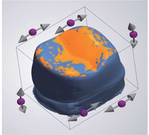
The maxillary first molar artificial tooth was selected as the reference tooth. The GMs were fabricated through a silicone impression of a reference tooth, and PMs were fabricated by a digital impression (n=9, in each group). The reference tooth and experimental models were scanned using a 3 shape convince(TM) scan system. Each GM and PM image was superimposed on the registered reference model (RM) and 2D images were obtained. The discrepancies of the points registered on the superimposed images were measured and defined as GM-RM group and PM-RM group. Statistical analysis was performed using a Student's T-test (alpha=0.05).
RESULTS
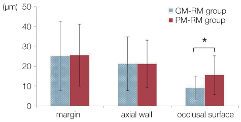
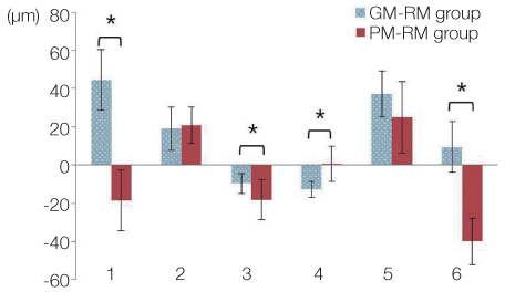
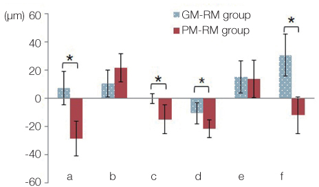
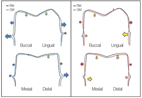
A comparison of the absolute value of the discrepancy revealed a significant difference between the two groups only at the occlusal surface. The GM group showed a smaller mean discrepancy than the PM group. Significant differences in the GM-RM group and PM-RM group were observed in the margins (point a and f), mesial mid-axial wall (point b) and occlusal surfaces (point c and d).
CONCLUSION
Under the conditions examined, the digitally fabricated polyurethane model showed a tendency for a reduced size in the margin than the reference tooth. The conventional gypsum model showed a smaller discrepancy on the occlusal surface than the polyurethane model.
MeSH Terms
Figure
Cited by 2 articles
-
Comparison of the accuracy of digital impressions and traditional impressions: Systematic review
Kyoung-Rok Kim, Kweonsoo Seo, Sunjai Kim
J Korean Acad Prosthodont. 2018;56(3):258-268. doi: 10.4047/jkap.2018.56.3.258.Accuracy and reproducibility of 3D digital tooth preparations made by gypsum materials of various colors
Fa-Bing Tan, Chao Wang, Hong-Wei Dai, Yu-Bo Fan, Jin-Lin Song
J Adv Prosthodont. 2018;10(1):8-17. doi: 10.4047/jap.2018.10.1.8.
Reference
-
1. Spear F, Puri S, Manji I. In-office CAD/CAM: the future of your practice? Dent Today. 2009; 28:6870–71.2. Presbyter T, Hawthorne JG, Smith CS. On divers arts: The foremost medieval treatise on painting, glassmaking and metalwork. Mineola, NY: Dover Publications;1979.3. Miyazaki T, Hotta Y, Kunii J, Kuriyama S, Tamaki Y. A review of dental CAD/CAM: current status and future perspectives from 20 years of experience. Dent Mater J. 2009; 28:44–56.4. Sutton AF, McCord JF. Variations in tooth preparations for resin-bonded all-ceramic crowns in general dental practice. Br Dent J. 2001; 191:677–681.5. Goodacre CJ, Campagni WV, Aquilino SA. Tooth preparations for complete crowns: an art form based on scientific principles. J Prosthet Dent. 2001; 85:363–376.6. Lowe RA. CAD/CAM Dentistry and Chariside Digital Impression Making. Accessed August 2012. www.ineedce.com.7. Mörmann WH. The evolution of the CEREC system. J Am Dent Assoc. 2006; 137:7S–13S.8. Allen KL, Schenkel AB, Estafan D. An overview of the CEREC 3D CAD/CAM system. Gen Dent. 2004; 52:234–235.9. Beuer F, Schweiger J, Edelhoff D. Digital dentistry: an overview of recent developments for CAD/CAM generated restorations. Br Dent J. 2008; 204:505–511.10. Henkel GL. A comparison of fixed prostheses generated from conventional vs digitally scanned dental impressions. Compend Contin Educ Dent. 2007; 28:422–424. 426–428. 430–431.11. Christensen GJ. Impressions are changing: deciding on conventional, digital or digital plus in-office milling. J Am Dent Assoc. 2009; 140:1301–1304.12. Galhano GÁ, Pellizzer EP, Mazaro JV. Optical impression systems for CAD-CAM restorations. J Craniofac Surg. 2012; 23:e575–e579.13. Touchstone A, Nieting T, Ulmer N. Digital transition: the collaboration between dentists and laboratory technicians on CAD/CAM restorations. J Am Dent Assoc. 2010; 141:15S–19S.14. Christensen GJ. The state of fixed prosthodontic impressions: room for improvement. J Am Dent Assoc. 2005; 136:343–346.15. Survey respondents are upbeat and optimistic about the state of our industry. Lab Manag Today. 2000; 16:9–15.16. Syrek A, Reich G, Ranftl D, Klein C, Cerny B, Brodesser J. Clinical evaluation of all-ceramic crowns fabricated from intraoral digital impressions based on the principle of active wavefront sampling. J Dent. 2010; 38:553–559.17. Ender A, Mehl A. Full arch scans: conventional versus digital impressions--an in-vitro study. Int J Comput Dent. 2011; 14:11–21.18. Reich S, Wichmann M, Nkenke E, Proeschel P. Clinical fit of all-ceramic three-unit fixed partial dentures, generated with three different CAD/CAM systems. Eur J Oral Sci. 2005; 113:174–179.19. Bindl A, Mörmann WH. Marginal and internal fit of all-ceramic CAD/CAM crown-copings on chamfer preparations. J Oral Rehabil. 2005; 32:441–447.20. Scotti R, Cardelli P, Baldissara P, Monaco C. Clinical fitting of CAD/CAM zirconia single crowns generated from digital intraoral impressions based on active wavefront sampling. J Dent. 2011; 10. 17.21. Wöstmann B, Rehmann P, Balkenhol M. Accuracy of impressions obtained with dual-arch trays. Int J Prosthodont. 2009; 22:158–160.22. Quick DC, Holtan JR, Ross GK. Use of a scanning laser three-dimensional digitizer to evaluate dimensional accuracy of dental impression materials. J Prosthet Dent. 1992; 68:229–235.23. Mehl A, Ender A, Mörmann W, Attin T. Accuracy testing of a new intraoral 3D camera. Int J Comput Dent. 2009; 12:11–28.24. Luthardt RG, Loos R, Quaas S. Accuracy of intraoral data acquisition in comparison to the conventional impression. Int J Comput Dent. 2005; 8:283–294.25. Huh JB, Kim US, Kim HY, Kim JE, Lee JY, Kim YS, Jeon YC, Shin SW. Marginal and internal fitness of three-unit zirconia cores fabricated using several CAD/CAM systems. J Korean Acad Prosthodont. 2011; 49:236–244.26. Lowe RA. Digital Master Impressions: A Clinical Reality. Accessed August 2012. URL: http://www.cadentinc.com/pdfs/itero_DC_08_09.pdf.27. van Noort R. The future of dental devices is digital. Dent Mater. 2012; 28:3–12.28. Miyazaki T, Hotta Y. CAD/CAM systems available for the fabrication of crown and bridge restorations. Aust Dent J. 2011; 56:97–106.29. Tinschert J, Natt G, Hassenpflug S, Spiekermann H. Status of current CAD/CAM technology in dental medicine. Int J Comput Dent. 2004; 7:25–45.30. Sorensen JA. A standardized method for determination of crown margin fidelity. J Prosthet Dent. 1990; 64:18–24.31. Moon BH, Yang JH, Lee SH, Chung HY. A study on the marginal fit of all-ceramic crown using CCD camera. J Korean Acad Prosthodont. 1998; 36:273–292.32. Tjan AH, Nemetz H, Nguyen LT, Contino R. Effect of tray space on the accuracy of monophasic polyvinylsiloxane impressions. J Prosthet Dent. 1992; 68:19–28.33. Jørgensen KD. Factors affecting the film thickness of Zinc phosphate cements. Acta Odontol Scand. 1960; 18:479–490.34. Christensen GJ. Marginal fit of gold inlay castings. J Prosthet Dent. 1966; 16:297–305.35. McLean JW, von Fraunhofer JA. The estimation of cement film thickness by an in vivo technique. Br Dent J. 1971; 131:107–111.36. McLean JW. Polycarboxylate cements. Five years' experience in general practice. Br Dent J. 1972; 132:9–15.
- Full Text Links
- Actions
-
Cited
- CITED
-
- Close
- Share
- Similar articles
-
- Comparison of the accuracy of implant digital impression coping
- Accuracy of dies fabricated by various three dimensional printing systems: a comparative study
- Accuracy of 14 intraoral scanners for the All-on-4 treatment concept: a comparative in vitro study
- Subtractive versus additive indirect manufacturing techniques of digitally designed partial dentures
- Accuracy and precision of polyurethane dental arch models fabricated using a three-dimensional subtractive rapid prototyping method with an intraoral scanning technique


