Accuracy and precision of polyurethane dental arch models fabricated using a three-dimensional subtractive rapid prototyping method with an intraoral scanning technique
- Affiliations
-
- 1Department of Dental Laboratory Science and Engineering, College of Health Science, Korea University, Seoul, Korea. kimhaey@korea.ac.kr
- 2Department of Public Health Sciences, Graduate School, Korea University, Seoul, Korea.
- 3BK21+ Program in Public Health Sciences, Korea University, Seoul, Korea.
- KMID: 2273429
- DOI: http://doi.org/10.4041/kjod.2014.44.2.69
Abstract
OBJECTIVE
This study aimed to evaluate the accuracy and precision of polyurethane (PUT) dental arch models fabricated using a three-dimensional (3D) subtractive rapid prototyping (RP) method with an intraoral scanning technique by comparing linear measurements obtained from PUT models and conventional plaster models.
METHODS
Ten plaster models were duplicated using a selected standard master model and conventional impression, and 10 PUT models were duplicated using the 3D subtractive RP technique with an oral scanner. Six linear measurements were evaluated in terms of x, y, and z-axes using a non-contact white light scanner. Accuracy was assessed using mean differences between two measurements, and precision was examined using four quantitative methods and the Bland-Altman graphical method. Repeatability was evaluated in terms of intra-examiner variability, and reproducibility was assessed in terms of inter-examiner and inter-method variability.
RESULTS
The mean difference between plaster models and PUT models ranged from 0.07 mm to 0.33 mm. Relative measurement errors ranged from 2.2% to 7.6% and intraclass correlation coefficients ranged from 0.93 to 0.96, when comparing plaster models and PUT models. The Bland-Altman plot showed good agreement.
CONCLUSIONS
The accuracy and precision of PUT dental models for evaluating the performance of oral scanner and subtractive RP technology was acceptable. Because of the recent improvements in block material and computerized numeric control milling machines, the subtractive RP method may be a good choice for dental arch models.
Figure
-
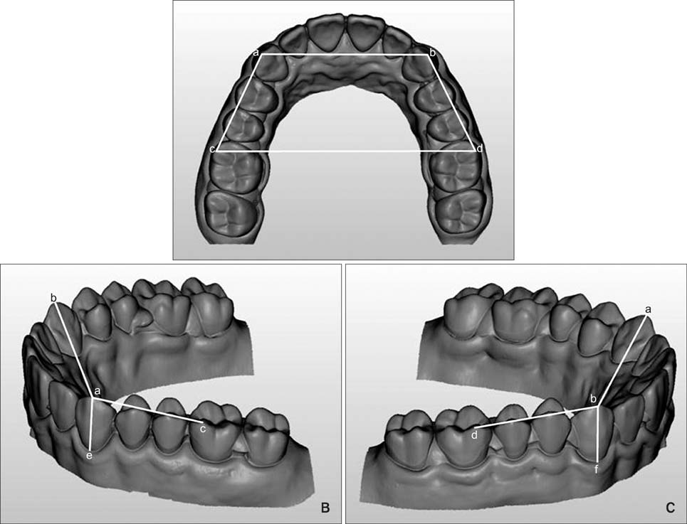
Figure 1 Reference points and linear measurements on the upper full arch model. A, Upper view; B, oblique view from the right side; C, oblique view from left side. See Table 1 for the definition of the reference points.

Figure 2 Bland-Altman plot. A, Repeatability, intra-examiner variability; B, reproducibility, inter-examiner variability; C, reproducibility, inter-method variability. '95% limit of agreement (LoA)' represents mean difference ± 1.96 standard deviation.
Cited by 3 articles
-
Comparison of occlusal contact areas of class I and class II molar relationships at finishing using three-dimensional digital models
Hyejoon Lee, Minji Kim, Youn-Sic Chun
Korean J Orthod. 2015;45(3):113-120. doi: 10.4041/kjod.2015.45.3.113.Accuracy of three-dimensional printing for manufacturing replica teeth
Keun-Young Lee, Jin-Woo Cho, Na-Young Chang, Jong-Moon Chae, Kyung-Hwa Kang, Sang-Cheol Kim, Jin-Hyoung Cho
Korean J Orthod. 2015;45(5):217-225. doi: 10.4041/kjod.2015.45.5.217.Implant prosthesis using intraoral scanner: Case Report
Byeong-Gil Kang, Hee-Jung Kim, Chae-Heon Chung
J Korean Acad Prosthodont. 2015;53(3):256-261. doi: 10.4047/jkap.2015.53.3.256.
Reference
-
1. Henkel S. A better first impression: manufacturing dental restorations using impressions. J Dent Technology. 2008; 13–16.2. Jones P. The iTero optical scanner for use with Invisalign: A descriptive review. Dent Implantol Update. 2008; 19:1–4.3. Barker TM, Earwaker WJ, Lisle DA. Accuracy of stereolithographic models of human anatomy. Australas Radiol. 1994; 38:106–111.
Article4. Kragskov J, Sindet-Pedersen S, Gyldensted C, Jensen KL. A comparison of three-dimensional computed tomography scans and stereolithographic models for evaluation of craniofacial anomalies. J Oral Maxillofac Surg. 1996; 54:402–411.
Article5. Cuperus AM, Harms MC, Rangel FA, Bronkhorst EM, Schols JG, Breuning KH. Dental models made with an intraoral scanner: a validation study. Am J Orthod Dentofacial Orthop. 2012; 142:308–313.6. Lill W, Solar P, Ulm C, Watzek G, Blahout R, Matejka M. Reproducibility of three-dimensional CT-assisted model production in the maxillofacial area. Br J Oral Maxillofac Surg. 1992; 30:233–236.
Article7. Santoro M, Galkin S, Teredesai M, Nicolay OF, Cangialosi TJ. Comparison of measurements made on digital and plaster models. Am J Orthod Dentofacial Orthop. 2003; 124:101–105.
Article8. Quimby ML, Vig KW, Rashid RG, Firestone AR. The accuracy and reliability of measurements made on computer-based digital models. Angle Orthod. 2004; 74:298–303.9. Zilberman O, Huggare JA, Parikakis KA. Evaluation of the validity of tooth size and arch width measurements using conventional and three-dimensional virtual orthodontic models. Angle Orthod. 2003; 73:301–306.10. Stevens DR, Flores-Mir C, Nebbe B, Raboud DW, Heo G, Major PW. Validity, reliability, and reproducibility of plaster vs digital study models: comparison of peer assessment rating and Bolton analysis and their constituent measurements. Am J Orthod Dentofacial Orthop. 2006; 129:794–803.
Article11. Watanabe-Kanno GA, Abrão J, Miasiro Junior H, Sánchez-Ayala A, Lagravère MO. Reproducibility, reliability and validity of measurements obtained from Cecile3 digital models. Braz Oral Res. 2009; 23:288–295.
Article12. Leifert MF, Leifert MM, Efstratiadis SS, Cangialosi TJ. Comparison of space analysis evaluations with digital models and plaster dental casts. Am J Orthod Dentofacial Orthop. 2009; 136:16.e1–16.e4.
Article13. Mullen SR, Martin CA, Ngan P, Gladwin M. Accuracy of space analysis with emodels and plaster models. Am J Orthod Dentofacial Orthop. 2007; 132:346–352.
Article14. Keating AP, Knox J, Bibb R, Zhurov AI. A comparison of plaster, digital and reconstructed study model accuracy. J Orthod. 2008; 35:191–201.
Article15. Creed B, Kau CH, English JD, Xia JJ, Lee RP. A comparison of the accuracy of linear measurements obtained from cone beam computerized tomography images and digital models. Semin Orthod. 2011; 17:49–56.
Article16. Alcan T, Ceylanoğlu C, Baysal B. The relationship between digital model accuracy and time-dependent deformation of alginate impressions. Angle Orthod. 2009; 79:30–36.
Article17. Dahlberg G. Statistical methods for medical and biological students. London: George Allen & Unwin Ltd.;1940. p. 122–132.18. Henriksen M, Lund H, Moe-Nilssen R, Bliddal H, Danneskiod-Samsøe B. Test-retest reliability of trunk accelerometric gait analysis. Gait Posture. 2004; 19:288–297.
Article19. Bland JM, Altman DG. Statistical methods for assessing agreement between two methods of clinical measurement. Lancet. 1986; 1:307–310.
Article20. Fleiss JL. The design and analysis of clinical experiments. New York: John Wiley & Sons;1986. p. 76–78.21. BeGole EA. Statistics for the orthodontist. In : Graber TM, Vanarsdall RL, editors. Orthodontics: current principles and techniques. 3rd ed. St. Louis: Mosby;2000. p. 339–352.22. Dalstra M, Melsen B. From alginate impressions to digital virtual models: accuracy and reproducibility. J Orthod. 2009; 36:36–41.
Article23. Mok KH, Cooke MS. Space analysis: a comparison between sonic digitization (DigiGraph Workstation) and the digital caliper. Eur J Orthod. 1998; 20:653–661.
Article24. Asquith J, Gillgrass T, Mossey P. Three-dimensional imaging of orthodontic models: a pilot study. Eur J Orthod. 2007; 29:517–522.
Article25. Linnet K. Limitations of the paired t-test for evaluation of method comparison data. Clin Chem. 1999; 45:314–315.
Article26. Donatelli RE, Lee SJ. How to report reliability in orthodontic research: Part 1. Am J Orthod Dentofacial Orthop. 2013; 144:156–161.
Article27. Reich S, Uhlen S, Gozdowski S, Lohbauer U. Measurement of cement thickness under lithium disilicate crowns using an impression material technique. Clin Oral Investig. 2011; 15:521–526.
Article28. Birnbaum NS, Aaronson HB. Dental impressions using 3D digital scanners: virtual becomes reality. Compend Contin Educ Dent. 2008; 29:494496498–505.29. Klein HM, Schneider W, Alzen G, Voy ED, Günther RW. Pediatric craniofacial surgery: comparison of milling and stereolithography for 3D model manufacturing. Pediatr Radiol. 1992; 22:458–460.
Article30. Petrzelka JE, Frank MC. Advanced process planning for subtractive rapid prototyping. Rapid Prototyping J. 2010; 16:216–224.
Article
- Full Text Links
-
- Actions
-
Cited
- CITED
-
- Close
- Share
-
- Similar articles
-
- A case report of a surgical guide fabricated via intraoral scanning-based implant planning and wax-based rapid prototyping
- A comparison of the precision of three-dimensional images acquired by 2 digital intraoral scanners: effects of tooth irregularity and scanning direction
- Accuracy of CAD-CAM RPD framework according to manufacturing method: A literature review
- A STUDY ON THE DIMENSIONAL ACCURACY OF MODELS USING 3-DIMENSIONAL COMPUTER TOMOGRAPHY AND 2 RAPID PROTOTYPING METHODS
- Comparison of intraoral scanning and conventional impression techniques using 3-dimensional superimposition
