J Korean Acad Conserv Dent.
2009 May;34(3):208-214. 10.5395/JKACD.2009.34.3.208.
Effects of condensation techniques and canal sizes on the microleakage of orthograde MTA apical plug in simulated canals
- Affiliations
-
- 1Department of Conservative Dentistry, School of Dentistry, Pusan National University, Busan, Korea. golddent@pusan.ac.kr
- KMID: 2176146
- DOI: http://doi.org/10.5395/JKACD.2009.34.3.208
Abstract
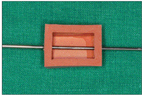
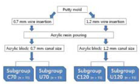
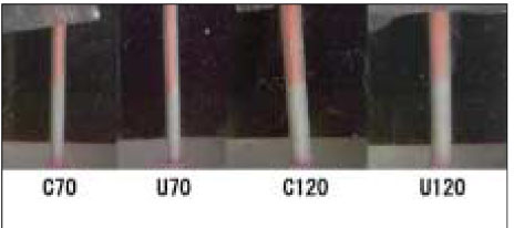
- The purpose of this study was to compare the dye leakage of MTA (mineral trioxide aggregate) apical plug produced by two orthograde placement techniques (hand condensation technique and ultrasonically assisted hand condensation technique). To simulate straight canal, 60 transparent acrylic blocks with straight canal were fabricated. These transparent acrylic blocks were divided into 2 groups (Group C; hand condensation technique (HC) and Group U; ultrasonically assisted hand condensation technique (UAHC)) of 30 blocks with each MTA application method. Each group was divided into 2 subgroups (n = 15) with different canal size of #70 (subgroup C70 and subgroup U70) and #120 (subgroup C120 and subgroup U120). After apical plug was created, a wet paper point was placed over the MTA plug and specimen was kept in a humid condition at room temperature to allow MTA to set. After 24 hours, remaining canal space was backfilled using Obtura II. All specimens were transferred to floral form socked by 0.2% rhodamine B solution and stored in 100% humidity at room temperature. After 48 hours, resin block specimens were washed and scanned using a scanner. The maximum length of microleakage was measured from the scanned images of four surfaces of each resin block using Photoshop 6.0. Statistical analysis was performed with Mann-Whitney U test. Group U of UAHC had significantly lower leakage than Group C of HC in #70-size canal (subgroup U70) (p < 0.05).
Keyword
MeSH Terms
Figure
Reference
-
1. Simon S, Rilliard F, Berdal A, Machtou P. The use of mineral trioxide aggregate in one-visit apexification treatment: a prospective study. Int Endod J. 2007. 40:186–197.
Article2. Pace R, Giuliani V, Pini Prato L, Baccetti T, Pagavino G. Apical plug technique using mineral trioxide aggregate: results from a case series. Int Endod J. 2007. 40:478–484.
Article3. Kwon JY, Lim SS, Beak SH, Bea KS, Kang MH, Lee WC. The effect of mineral trioxide aggregate on the production of growth factors and cytokine by human periodontal ligament fibroblasts. J Korean Acad Conserv Dent. 2007. 32(3):191–197.
Article4. Yun YR, Yang IS, Hwang YC, Hwang IN, Choi HR, Yoon SJ, Kim SH, Oh WM. Pulp response of mineral trioxide aggregate, calcium sulfate or calcium hydroxide. J Korean Acad Conserv Dent. 2007. 32(2):95–101.
Article5. Rafter M. Apexification: a review. Dent Traumatol. 2005. 21(1):1–8.
Article6. Shabahang S, Torabinejad M, Boyne PP, Abedi H, McMillan P. A comparative study of root-end induction using osteogenic protein-1, calcium hydroxide, and mineral trioxide aggregate in dogs. J Endod. 1999. 25(1):1–5.
Article7. Andreasen JO, Farik B, Munksgaard EC. Long-term calcium hydroxide as a root canal dressing may increase risk of root fracture. Dent Traumatol. 2002. 18(3):134–137.
Article8. Torabinejad M, Chivian N. Clinical applications of mineral trioxide aggregate. J Endod. 1999. 25(3):197–205.
Article9. D'Arcangelo C, D'Amario M. Use of MTA for orthograde obturation of nonvital teeth with open apices: report of two cases. Oral Surg Oral Med Oral Pathol Oral Radiol Endod. 2007. 104(4):e98–e101.10. Al-Kahtani A, Shostad S, Schifferle R, Bhambhani S. In-vitro evaluation of microleakage of an orthograde apical plug of mineral trioxide aggregate in permanent teeth with simulated immature apices. J Endod. 2005. 31(2):117–119.
Article11. Roberts HW, Toth JM, Berzins DW, Charlton DG. Mineral trioxide aggregate material use in endodontic treatment: a review of the literature. Dent Mater. 2007. 24(2):149–164.
Article12. Lawley GR, Schindler WG, Walker WA 3rd, Kolodrubetz D. Evaluation of ultrasonically placed MTA and fracture resistance with intracanal composite resin in a model of apexification. J Endod. 2004. 30(3):167–172.
Article13. Giuliani V, Baccetti T, Pace R, Pagavino G. The use of MTA in teeth with necrotic pulps and open apices. Dent Traumatol. 2002. 18(4):217–221.14. Martin RL, Monticelli F, Brackett WW, Loushine RJ, Rockman RA, Ferrari M, Pashley DH, Tay FR. Sealing properties of mineral trioxide aggregate orthograde apical plugs and root fillings in an in vitro apexification model. J Endod. 2007. 33(3):272–275.
Article15. Ghaziani P, Aghasizadeh N, Sheikh-Nezami M. Endodontic treatment with MTA apical plugs: a case report. J Oral Sci. 2007. 49(4):325–329.
Article16. Nekoofar MH, Adusei G, Sheykhrezae MS, Hayes SJ, Bryant ST, Dummer PM. The effect of condensation pressure on selected physical properties of mineral trioxide aggregate. Int Endod J. 2007. 40(6):453–461.
Article17. Yeung P, Liewehr FR, Moon PC. A quantitative comparison of the fill density of MTA produced by two placement techniques. J Endod. 2006. 32(5):456–459.
Article18. Aminoshariae A, Hartwell GR, Moon PC. Placement of mineral trioxide aggregate using two different techniques. J Endod. 2003. 29(10):679–682.
Article19. Hachmeister DR, Schindler WG, Walker WA 3rd, Thomas DD. The sealing ability and retention characteristics of mineral trioxide aggregate in a model of apexification. J Endod. 2002. 28(5):386–390.
Article20. Torabinejad M, Hong CU, McDonald F, Pitt Ford TR. Physical and chemical properties of a new root-end filling material. J Endod. 1995. 21(7):349–353.
Article21. Chang SW, Yoo HM, Park DS, Oh TS, Bae KS. Ingredients and cytotoxicity of MTA and 3 kinds of Portland cement. J Korean Acad Conserv Dent. 2008. 33(4):369–376.
Article22. Dammaschke T, Gerth HU, Züchner H, Schäfer E. Chemical and physical surface and bulk material characterization of white ProRoot MTA and two Portland cements. Dent Mater. 2005. 21(8):731–738.
Article23. Stock CJ. Current status of the use of ultrasound in endodontics. Int Dent J. 1991. 41(3):175–182.24. Plotino G, Pameijer CH, Grande NM, Somma F. Ultrasonics in endodontics: a review of the literature. J Endod. 2007. 33(2):81–95.
Article25. Newatia S. Concrete vibrators. [Web document]. Aurtech Global. Accessed May 12, 2008. http://www.aurtechindia.com/convib.htm.26. Aqrabawi J. Sealing ability of amalgam, super EBA cement, and MTA when used as retrograde filling materials. Br Dent J. 2000. 11. 188(5):266–268.
Article27. Tanomaru Filho M, Figueiredo FA, Tanomaru JM. Effect of different dye solutions on the evaluation of the sealing ability of Mineral Trioxide Aggregate. Braz Oral Res. 2005. 19(2):119–122.
Article28. Torabinejad M, Watson TF, Pitt Ford TR. Sealing ability of a mineral trioxide aggregate when used as a root end filling material. J Endod. 1993. 19(12):591–515.
Article29. Kontakiotis EG, Wu MK, Wesselink PR. Effect of calcium hydroxide dressing on seal of permanent root filling. Endod Dent Traumatol. 1997. 13(6):281–284.
Article30. Montesin FE, Curtis RV, Ford TR. Characterization of Portland cement for use as a dental restorative material. Dent Mater. 2005. 22(6):569–575.
Article
- Full Text Links
- Actions
-
Cited
- CITED
-
- Close
- Share
- Similar articles
-
- Failure of orthograde MTA filling: MTA wash-out?
- Comparison of warm gutta-percha condensation techniques in ribbon shaped canal: weight of filled gutta-percha
- Evaluation of the influence of apical sizes on the apical sealing ability of the modified continuous wave technique
- Effectiveness of customized master cone on apical sealing in various apical size of prepared root canals
- Microleakage of resilon: Effects of several self-etching primer

