Effectiveness of customized master cone on apical sealing in various apical size of prepared root canals
- Affiliations
-
- 1Department of Conservative Dentistry, College of Dentistry, KyungHee University, Korea.
- KMID: 2175549
- DOI: http://doi.org/10.5395/JKACD.2002.27.1.066
Abstract
- The purpose of this study is to evaluate the effectiveness of customized master cone on apical sealing in various apical size of prepared root canals, that is MAF(Master Apical File) and to know at which apical size the apical leakage is to be significantly reduced using customized master cone. 120 extracted single rooted premolars were divided into four groups according to their apical size(MAF), #30, 40, 50 and 60. And then, each group was subdivided into three in accordance with three obturation methods, lateral condensation with standardized master cone, lateral condensation with chloroform-dipped customized master cone, and continuous wave of obturation technique. Resorcinol-formaldehyde resin was used for the microleakage test of this study. Teeth were sectioned horizontally at 1.5mm(Level 1), 2.5mm(Level 2), and 3.5mm(Level 3) from the anatomical root apex using low speed microtome. All sections were examined under x40 magnification with a stereomicroscope, photographed, and then scanned. With the scanned images, resin-infiltrated area presenting the microleakage was calculated using SigmaScan/Image, and the ratio of leakage to the total root canal area of each group was analyzed statistically(one way ANOVA). The results were as follows; 1. In groups of MAF #30, there was no significant difference of mean leakage ratio among three obturation methods at all three levels. 2. In groups of MAF #40, the group using lateral condensation with customized master cone had the lowest mean leakage ratio at all three levels, but there was no significant difference among three obturation techniques. 3. In groups of MAF #50, the mean leakage ratio of the group using lateral condensation with standard master cone was the highest among those of three obturation techniques at level 1, and this difference was statistically significant(p<0.05). 4. In groups of MAF #60, the groups using lateral condensation with standard master cone had also the highest mean leakage ratio at all levels, but there was no significant difference at level 1 and 2. At level 3, the leakage of the group using lateral condensation with standard master cone was significantly higher than that of the group using continuous wave of obturation(p<0.05). The results of this study suggested that the obturation method using customized master cone or the continuous wave of obturation is more effective for apical sealing than that using standardized master cone when MAF is larger than #50.
Figure
-

Fig. 1 Mean leakage ratio(%) at 3 Levels (MAF #30) A: Lateral condensation with standard master cone B: Lateral condensation with customized master cone C: Continuous wave of obturation

Fig. 2 Mean leakage ratio(%) at 3 Levels (MAF #40) A: Lateral condensation with standard master cone B: Lateral condensation with customized master cone C: Continuous wave of obturation
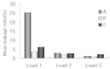
Fig. 3 Mean leakage ratio(%) at 3 Levels (MAF #50) A: Lateral condensation with standard master cone B: Lateral condensation with customized master cone C: Continuous wave of obturation
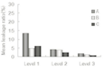
Fig. 4 Mean leakage ratio(%) at 3 Levels (MAF #60) A: Lateral condensation with standard master cone B: Lateral condensation with customized master cone C: Continuous wave of obturation

Fig. 5 Representative photograph of the group using lateral condensation with standard master cone at MAF #30, Level 1 (×40).

Fig. 6 Representative photograph of the group using lateral condensation with customized master cone at MAF #30, Level 1 (×40).
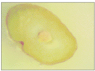
Fig. 7 Representative photograph of the group using continuous wave of obturation at MAF #30, Level 1 (× 40).

Fig. 8 Representative photograph of the group using lateral condensation with standard master cone at MAF #40, Level 1 (×40).
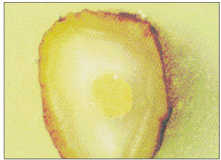
Fig. 9 Representative photograph of the group using lateral condensation with customized master cone at MAF #40, Level 1 (×40).
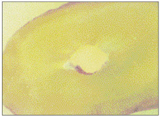
Fig. 10 Representative photograph of the group using continuous wave of obturation at MAF #40, Level 1 (× 40).
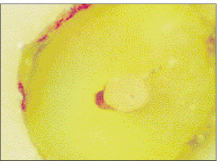
Fig. 11 Representative photograph of the group using lateral condensation with standard master cone at MAF #50, Level 1 (×40).

Fig. 12 Representative photograph of the group using lateral condensation with master cone at MAF #50, Level 1 (×40).
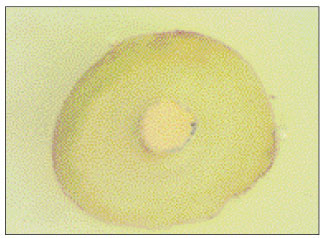
Fig. 13 Representative photograph of the group using continuous wave of obturation at MAF #50, Level 1 (× 40).

Fig. 14 Representative photograph of the group using lateral condensation with standard master cone at MAF #60, Level 1 (×40).

Fig. 15 Representative photograph of the group using lateral condensation with customized master cone at MAF #60, Level 1 (×40).
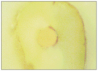
Fig. 16 Representative photograph of the group using continuous wave of obturation at MAF #60, Level 1 (× 40).
Cited by 1 articles
-
Influence of plugger penetration depth on the apical extrusion of root canal sealer in Continuous Wave of Condensation Technique
Ho-Young So, Young-Mi Lee, Kwang-Keun Kim, Ki-Ok Kim, Young-Kyung Kim, Sung-Kyo Kim
J Korean Acad Conserv Dent. 2004;29(5):439-445. doi: 10.5395/JKACD.2004.29.5.439.
Reference
-
1. Schilder H. Cleaning and shaping the root canal. Dent Clin North Am. 1974. 18:269.2. Dow PR, Ingle JI. Isotope determination of root canal failure. Oral Surg Oral Med Oral Pathol. 1955. 8:1100–1104.
Article3. Wein FS. Endodontic Therapy. 1996. 5th ed. Mosby;14317435–436.4. Callahan JR. Rosin solution for the sealing of the dentinal tubuli and as an adjuvant in filling the root canals. J Allied Dent Soc. 1914. 9:53.5. Wong M, Peters DD, Lorton L, Bernier EN. Comparison of gutta-percha filling techniques: three chloroform gutta-percha filling techniques Part 2. J Endod. 1982. 8:4–9.
Article6. Morse DR, Martell B, Pike GC, Fantasia J, Esposito JV, Furst LM. A Comparative Tissue Toxicity Evaluation of Gutta-Percha Root Canal Sealers. Part I. Six-hour Findings. J Endod. 1984. 10:246–249.
Article7. Barbosa SV, Burkard DH, Spångberg LSW. Cytotoxic Effects of Gutta-percha Solvents. J Endod. 1994. 20:6–8.
Article8. Beatty RG, Zakariasen KL. Apical leakage associated with three obturation techniques in large and small root canals. Int Endod J. 1984. 17:67–72.
Article9. Gutmann JL, Witherspoon DE. Cohen S, Burns RC, editors. Obturation of the Cleaned and Shaped Root Canal System. Pathways of the pulp. 1998. 7th ed. Mosby;258–259. 32710. Buchanan LS. The continuous wave of obturation technique: 'Centered' condensation of warm gutta-percha in 12 seconds. Dent Today. 1996. Jan. 15:60–62. 64–67.11. Narracott P. An in vitro comparison of the single cone and lateral condensation techniques using 'friction-fitted' and 'solvent dip-fitted' primary gutta-percha cones. Aust Dent J. 1989. 34:49–51.
Article12. Metzger Z, Nissan R, Tagger M, Tamse A. Apical Seal by Customized versus Standardized Master Cones: A Comparative Study in Flat and Round Canals. J Endod. 1988. 14:381–384.
Article13. Christie WH, Peikoff MD. Direct impression technique; Sealing prepared apical foramen. J Can Dent Assoc. 1980. 3:174–180.14. Zakariasen KL, Stadem PS. Microleakage associated with modified eucapercha and chloropercha root-canal-filling techniques. Int Endod J. 1982. 15:67–70.
Article15. Keane KM, Harrington GW. The Use of Chloroform-softened Gutta-percha Master Cone and Its Effect on the Apical Seal. J Endod. 1984. 10:57–63.
Article16. Metzger Z, Assif O, Tamse A. Residual Chloroform and Plasticity in Customized Gutta-percha Master Cones. J Endod. 1998. 14:546–549.
Article17. West JD, Roane JB. Cohen S, Burns RC, editors. Cleaning and Shaping the Root Canal System. Pathways of the pulp. 1998. 7th ed. Mosby;232.18. Luiten DJ, Morgan LA, Baumgartner JC, Marshall JG. A Comparison of Four Instrumentation Techniques on Apical Canal Transportation. J Endod. 1995. 21:26–32.
Article19. Christie WH, Peikoff MD. Conservative treatment of apical foramen; New root canal techniques. J Can Dent Assoc. 1980. 46:183–188.20. Shin JH, Choi HY, Park SJ, Choi KK, Choi KW. A study on the apical sealing of obturation techniques in large apical foramen. Kyung Hee Dent J. 1999. 21:1–16.21. Ingle JI, West JD. Ingle JI, Bakland LK, editors. Obturation of the radicular space. Endodontics. 1994. 4th ed. A Lea & Febiger Book;266.22. Schilder H. Filling root canals in three dimensions. Dent Clin North Am. 1967. 11:723–744.
Article23. Jerome CE. Warm Vertical Gutta-percha Obturation: A Technique Update. J Endod. 1994. 20:97–99.
Article
- Full Text Links
-
- Actions
-
Cited
- CITED
-
- Close
- Share
-
- Similar articles
-
- Obturation efficiency of non-standardized gutta-percha cone in curved root canals prepared with 0.06 taper nickel-titanium instruments
- Apical prepration size in infected root canals
- A comparison of master apical file size according to instrumentation in type II root canal
- Evaluation of the influence of apical sizes on the apical sealing ability of the modified continuous wave technique
- Bacterial leakage and micro-computed tomography evaluation in round-shaped canals obturated with bioceramic cone and sealer using matched single cone technique
