Comparison of biocompatibility of four root perforation repair materials
- Affiliations
-
- 1Department of Conservative Dentistry, School of Dentistry, Chonnam National University, Korea. wmoh@chonnam.ac.kr
- 2Department of Pharmacology and Dental Therapeutics, School of Dentistry, Chonnam National University, Korea.
- 3DSRI, Chonnam National University, Korea.
- 42nd stage of BK21, Chonnam National University, Korea.
- KMID: 2176144
- DOI: http://doi.org/10.5395/JKACD.2009.34.3.192
Abstract
- This study was carried out in order to determine in vitro biocompatibility of white mineral trioxide aggregate (MTA), and to compare it with that of the commonly used materials, i. e. calcium hydroxide liner (Dycal), glass ionomer cement (GIC), and Portland cement which has a similar composition of MTA. To assess the biocompatibility of each material, cytotoxicity was examined using MG-63 cells. The degree of cytotoxicity was evaluated by scanning electron microscopy (SEM) and a colorimetric method, based on reduction of the tetrazolium salt 2,3 bis {2methoxy 4nitro 5[(sulfenylamino) carbonyl] 2H tetrazolium hydroxide} (XTT) assay. The results of SEM revealed the cells in contact with GIC, MTA, and Portland cement at 1 and 3 days were apparently healthy. In contrast, cells in the presence of Dycal appeared rounded and detached. In XTT assay, the cellular activities of the cells incubated with all the test materials except Dycal were similar, which corresponded with the SEM observation. The present study supports the view that MTA is a very biocompatible root perforation repair material. It also suggests that cellular response of Portland cement and GIC are very similar to that of MTA.
Keyword
MeSH Terms
-
Acrylic Resins
Aluminum Compounds
Calcium Compounds
Calcium Hydroxide
Drug Combinations
Glass Ionomer Cements
Glutamates
Guanine
Humans
Hydroxides
Microscopy, Electron, Scanning
Minerals
Oxides
Silicates
Silicon Dioxide
Pemetrexed
Acrylic Resins
Aluminum Compounds
Calcium Compounds
Calcium Hydroxide
Drug Combinations
Glass Ionomer Cements
Glutamates
Guanine
Hydroxides
Minerals
Oxides
Silicates
Silicon Dioxide
Figure
-

Figure 1 Cells after the incubation with test material for 1 day (×250, a: Dycal, b: GIC, c: MTA, d: Portland cement)
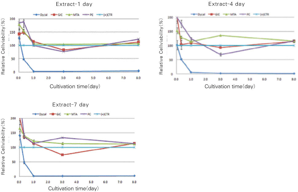
Figure 2 Cell viability after 1 day, 3 day, 8 day incubation with the extract from Dycal, GIC, MTA, and Portland cement as measured by XTT assay (a: 1 day extract, b: 4 day extract, C: 7 day extract), (+)CTR : Positive control) *Significantly difference (p < 0.05) between the materials.
Cited by 3 articles
-
The effect of several root-end filling materials on MG63 osteoblast-like cells
Jeong-Ho Lee, Won-Jun Shon, WooCheol Lee, Seung-Ho Baek
J Korean Acad Conserv Dent. 2010;35(3):222-228. doi: 10.5395/JKACD.2010.35.3.222.Physical and chemical properties of experimental mixture of mineral trioxide aggregate and glass ionomer cement
Yu-Na Jeong, So-Young Yang, Bum-Jun Park, Yeong-Joon Park, Yun-Chan Hwang, In-Nam Hwang, Won-Mann Oh
J Korean Acad Conserv Dent. 2010;35(5):344-352. doi: 10.5395/JKACD.2010.35.5.344.Biocompatibility of experimental mixture of mineral trioxide aggregate and glass ionomer cement
Min-Jae Oh, Yu-Na Jeong, In-Ho Bae, So-Young Yang, Bum-Jun Park, Jeong-Tae Koh, Yun-Chan Hwang, In-Nam Hwang, Won-Mann Oh
J Korean Acad Conserv Dent. 2010;35(5):359-367. doi: 10.5395/JKACD.2010.35.5.359.
Reference
-
1. Vajrabhaya LO, Korsuwannawong S, Jantarat J, Korre S. Biocompatibility of furcal perforation repair material using cell culture technique: Ketac Molar versus ProRoot MTA. Oral Surg Oral Med Oral Pathol Oral Radiol Endod. 2006. 102:e48–e50.
Article2. Souza NJ, Justo GZ, Oliveira CR, Haun M, Bincoletto C. Cytotoxicity of materials used in perforation repair tested using the V79 fibroblast cell line and the granulocyte-macrophage progenitor cells. Int Endod J. 2006. 39:40–47.
Article3. Tziafas D, Economides N. Formation of crystals on the surface of calcium hydroxide-containing materials In vitro. J Endod. 1999. 25:539–542.
Article4. Makkawy HA, Koka S, Lavin MT, Ewoldsen NO. Cytotoxicity of root perforation repair materials. J Endod. 1998. 24:477–479.
Article5. de Souza Costa CA, Hebling J, Garcia-Godoy F, Hanks CT. In vitro cytotoxicity of glass-ionomer cements. Biomaterials. 2003. 24:3853–3858.6. Yun YR, Yang IS, Hwang YC, Hwang IN, Choi HR, Yoon SJ, Kim SH, Oh WM. Pulp response of Mineral trioxide aggregate, calcium sulfate or calcium hydroxide. J Korean Acad Conserv Dent. 2007. 32:95–101.
Article7. Gorduysus M, Avcu N, Gorduysus O, Pekel A, Baran Y, Avcu F, Ural AU. Cytotoxic effects of four different endodontic materials in human periodontal ligament fibroblasts. J Endod. 2007. 33:1450–1454.
Article8. Chang SW, Yoo HM, Park DS, Oh TS, Bae KS. Ingredients and cytotoxicity of MTA and 3 kinds of Portland cements. J Korean Acad Conserv Dent. 2008. 33:369–376.
Article9. AL-Rabeah E, Perinpanayagam H, MacFarland D. Human alveolar bone cells interact with ProRoot and tooth-colored MTA. J Endod. 2006. 32:872–875.
Article10. Min KS, Kim HI, Park HJ, Pi SH, Hong CU, Kim EC. Human pulp cells response to Portland cement in Vitro. J Endod. 2007. 33:163–166.
Article11. Funteas UR, Wallace JA, Fochtman EW. A comparative analysis of mineral trioxide aggregate and Portland cement. Aust Endod J. 2003. 29:43–44.
Article12. Saidon J, He J, Zhu Q, Safavi K, Spångberg LS. Cell and tissue reactions to mineral trioxide aggregate and Portland cement. Oral Surg Oral Med Oral Pathol Oral Radiol Endod. 2003. 95:483–489.
Article13. Camilleri J, Montesin FE, Di Silvio L, Pitt Ford TR. The chemical constitution and biocompatibility of accelerated Portland cement for endodontic use. Int Endod J. 2005. 38:834–842.
Article14. Kim HJ, Baek SH, Bae KS. Cytotoxicity and genotoxicity of newly developed calcium phosphate-based root canal sealers. J Korean Acad Conserv Dent. 2006. 31:36–49.
Article15. Saw TY, Cao T, Yap AU, Lee Ng MM. Tooth slice organ culture and established cell line culture models for cytotoxicity assessment of dental materials. Toxicol in Vitro. 2005. 19:145–154.
Article16. Koh ET, McDonald F, Pitt Ford TR, Torabinejad M. Cellular response to Mineral Trioxide Aggregate. J Endod. 1998. 24:543–547.
Article17. Zhu Q, Haglund R, Safavi KE, Spangberg LS. Adhesion of human osteoblasts on root-end filling materials. J Endod. 2000. 26:404–406.
Article18. Mitchell PJ, Pitt Ford TR, Torabinejad M, McDonald F. Osteoblast biocompatibility of mineral trioxide aggregate. Biomaterials. 1999. 20:167–173.
Article19. Balto HA. Attachment and morphological behavior of human periodontal ligament fibroblasts to mineral trioxide aggregate: a scanning electron microscope study. J Endod. 2004. 30:25–29.
Article20. Torabinejad M, Hong CU, Pitt Ford TR, Kettering JD. Cytotoxicity of four root end filling materials. J Endod. 1995. 21:489–492.
Article21. Camilleri J, Montesin FE, Papaioannou S, McDonald F, Pitt Ford TR. Biocompatibility of two commercial forms of mineral trioxide aggregate. Int Endod J. 2004. 37:699–704.
Article22. Camilleri J, Montesin FE, Brady K, Sweeney R, Curtis RV, Ford TR. The constitution of mineral trioxide aggregate. Dent Mater. 2005. 21:297–303.
Article23. Scudiero DA, Shoemaker RH, Paull KD, Monks A, Tierney S, Nofziger TH, Currens MJ, Seniff D, Boyd MR. Evaluation of a soluble tetrazolium/formazan assay for cell growth and drug sensitivity in culture using human and other tumor cell lines. Cancer Res. 1988. 48:4827–4833.24. Leonardo RT, Consolaro A, Carlos IZ, Leonardo MR. Evaluation of cell culture cytotoxicity of five root canal sealers. J Endod. 2000. 26:328–330.
Article25. Scarano A, Manzon L, Di Giorgio R, Orsini G, Tripodi D, Piattelli A. Direct capping with four different materials in humans: histological analysis of odontoblast activity. J Endod. 2003. 29:729–734.
Article26. Abdullah D, Ford TR, Papaioannou S, Nicholson J, McDonald F. An evaluation of accelerated Portland cement as a restorative material. Biomaterials. 2002. 23:4001–4010.
Article
- Full Text Links
-
- Actions
-
Cited
- CITED
-
- Close
- Share
-
- Similar articles
-
- Biocompatibility of root-end filling materials: recent update
- A Case of Septal Perforation Repair with Middle Turbinate Flap
- Treatment options for iatrogenic root perforation: report of three cases
- Endonasal Septal Perforation Repair: Free Mucosal Graft With Lyoplant® Bioscaffold
- Pull-Out repair of the radial posterior horn tear near the root of the medial meniscus: Technical Note
