Ann Dermatol.
2008 Jun;20(2):82-85. 10.5021/ad.2008.20.2.82.
A Case of Disseminated Nocardiosis Secondary to the Skin Nodules in an Elderly Woman
- Affiliations
-
- 1Department of Dermatology, Inha University College of Medicine, Incheon, Korea. garden@inha.ac.kr
- 2Department of Internal Medicine, Inha University College of Medicine, Incheon, Korea.
- KMID: 2172090
- DOI: http://doi.org/10.5021/ad.2008.20.2.82
Abstract
- Nocardiosis refers to a locally invasive or disseminated infection associated with the Nocardia species. Most infections enter through the respiratory tract and then disseminate systemically. Rarely can a primary nocardial infection of the skin spread to contiguous structures or disseminate to other internal organs in immunocompromised hosts. We describe a 70-year-old woman who suffered from recurrent nodular skin lesions on her right hand, forearm and elbow following inoculation of a traumatic injury. Analysis of the purulent exudates obtained from the nodule revealed Nocardia species. After 20 days, a chest X-ray showed newly developed multiple nodules in both lungs. The diagnosis of systemic nocardiosis was established, and we treated this case with trimethoprim-sulfamethoxazole.
Keyword
MeSH Terms
Figure
-

Fig. 1 Multiple erythematous nodules on the right forearm (A) and the dorsum of hand (B).

Fig. 2 Granulomatous inflammation with central suppurative inflammation (A: H&E, ×40; B: H&E, ×400).
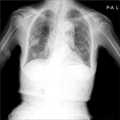
Fig. 3 Newly developed multiple nodules in both lungs.
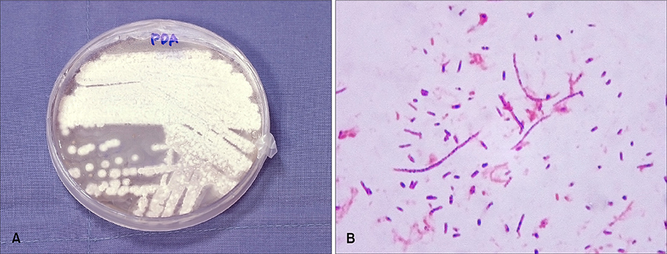
Fig. 4 (A) Whitish, wrinkled, heaped up colonies on potato dextrose agar plate cultured for 43 days. (B) Gram stain showed gram-positive branching organisms (×100).
Cited by 1 articles
-
Primary Cutaneous Nocardiosis Caused by Nocardia takedensis
Taek Geun Lee, Woo Jung Jin, Woo Seok Jeong, Seung Hyun Moon, Tae Gwang Kwon, Sook Kyung Lee, Hye Sook Kang, Hyun Hwangbo
Ann Dermatol. 2017;29(4):471-475. doi: 10.5021/ad.2017.29.4.471.
Reference
-
1. Kalb RE, Kaplan MH, Grossman ME. Cutaneous nocardiosis. Case reports and review. J Am Acad Dermatol. 1985; 13:125–133.2. Karakayali G, Karaarslan A, Artuz F, Alli N, Tekeli A. Primary cutaneous Nocardia asteroides. Br J Dermatol. 1998; 139:919–920.3. Lee SH, Suh CW, Choi JH, Sung KJ. A case of primary cutaneous sporotrichoid nocardiosis caused by Nocardia asteroides. Ann Dermatol. 1999; 11:90–93.
Article4. Aydingoz IE, Candan I, Dervent B, Hitit G. Primary cutaneous nocardiosis associated with intra-articular corticosteroid injection. Int J Dermatol. 2001; 40:196–198.
Article5. George SJ, Rivera AM, Hsu S. Disseminated cutaneous nocardiosis mimicking cellulitis and erythema nodosum. Dermatol Online J. 2006; 12:13.
Article6. Paredes BE, Hunger RE, Brassthen LR, Brand CU. Cutaneous nocardiosis caused by Nocardia brasiliensis after an insect bite. Dermatology. 1999; 198:159–161.
Article7. Wlodaver CG, Tolomeo T, Benear JB II. Primary cutaneous nocardiosis mimicking sporotrichosis. Arch Dermatol. 1988; 124:659–660.
Article8. Boixeda P, Espana A, Suarez J, Buzon L, Ledo A. Cutaneous nocardiosis and human immunodeficiency virus infection. Int J Dermatol. 1991; 30:804–805.
Article9. Kahn FW, Gornick CC, Tofte RW. Primary cutaneous Nocardia asteroides infection with dissemination. Am J Med. 1981; 70:859–863.
- Full Text Links
-
- Actions
-
Cited
- CITED
-
- Close
- Share
-
- Similar articles
-
- A Case of Disseminated Nocardiosis by Nocardia brasiliensis after Steroid Injection
- Disseminated nocardiosis including cerebral nocardiosis caused by Nocardia farcinica: a case report
- A Case of Disseminated Nocardiosis in Kidney Transplant Recipient
- Comparative Analysis of CT Findings and Clinical Outcomes in Adult Patients With Disseminated and Localized Pulmonary Nocardiosis
- A Case of Nocardia asteroides Isolated from Subcutaneous Abscess in a Pneumonic Patient with a Rejected Transplant Kidney
