J Neurocrit Care.
2023 Dec;16(2):107-110. 10.18700/jnc.230035.
Disseminated nocardiosis including cerebral nocardiosis caused by Nocardia farcinica: a case report
- Affiliations
-
- 1Department of Neurology, College of Medicine, Kyung Hee University, Seoul, Korea
- 2Division of Infectious Diseases, Department of Internal Medicine, College of Medicine, Kyung Hee University, Seoul, Korea
- 3Department of Laboratory Medicine, College of Medicine, Kyung Hee University, Seoul, Korea
- KMID: 2549488
- DOI: http://doi.org/10.18700/jnc.230035
Abstract
- Background
As nocardiosis presents with nonspecific symptoms, its diagnosis is often difficult, resulting in high mortality. Here, we report a rare case of disseminated Nocardia farcinica infection that was successfully managed with a combination of antibiotic therapies.
Case report
A 71-year-old man with idiopathic thrombocytopenia purpura presented with sudden-onset motor aphasia, motor weakness of the right limb, and fever. Brain magnetic resonance imaging revealed multiple scattered diffusion-restricted lesions. Multiple cutaneous and peritoneal nodules were also identified. We performed full-length 16S ribosomal ribonucleic acid (rRNA) sequencing and obtained basic local alignment search tool results against the National Center for Biotechnology Information 16S rRNA database, which showed the best match to be N. farcinica. After maintaining the antibiotics for approximately 56 days, the patient recovered full consciousness, and the motor weakness of the right limb improved.
Conclusion
Timely diagnosis of disseminated or cerebral nocardiosis and treatment with appropriate antibiotics are crucial for a desirable clinical outcome.
Keyword
Figure
-
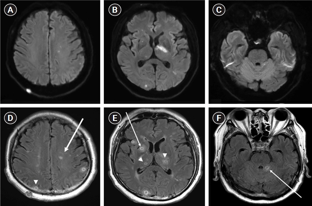
Fig. 1. Magnetic resonance imaging of the patient. (A-C) Multiple scattered diffusion-restricted lesions in both hemispheres, thalami, cerebella, and right basal ganglia are observed on the initial diffusion-weighted image. (D-F) Rim-enhancing lesions (arrows and arrowheads) with associated edema on T1 weighted imaging and a dark signal on T2 weighted imaging and consistent wall thickness suggest a brain abscess.
Reference
-
1. Curry WA. Human nocardiosis: a clinical review with selected case reports. Arch Intern Med. 1980; 140:818–26.2. Corti ME, Villafañe-Fioti MF. Nocardiosis: a review. Int J Infect Dis. 2003; 7:243–50.3. Lai CC, Lee LN, Teng LJ, Wu MS, Tsai JC, Hsueh PR. Disseminated Nocardia farcinica infection in a uraemia patient with idiopathic thrombocytopenia purpura receiving steroid therapy. J Med Microbiol. 2005; 54(Pt 11):1107–10.4. Wang H, Zhu Y, Cui Q, Wu W, Li G, Chen D, et al. Epidemiology and antimicrobial resistance profiles of the Nocardia species in China, 2009 to 2021. Microbiol Spectr. 2022; 10:e0156021.5. Malincarne L, Marroni M, Farina C, Camanni G, Valente M, Belfiori B, et al. Primary brain abscess with Nocardia farcinica in an immunocompetent patient. Clin Neurol Neurosurg. 2002; 104:132–5.6. de Montmollin E, Corcos O, Noussair L, Leflon-Guibout V, Belmatoug N, Joly F, et al. Retroperitoneal abscesses due to Nocardia farcinica: report of two cases in patients with malnutrition. Infection. 2012; 40:93–6.7. Verroken A, Janssens M, Berhin C, Bogaerts P, Huang TD, Wauters G, et al. Evaluation of matrix-assisted laser desorption ionization-time of flight mass spectrometry for identification of nocardia species. J Clin Microbiol. 2010; 48:4015–21.8. Khot PD, Bird BA, Durrant RJ, Fisher MA. Identification of Nocardia species by matrix-assisted laser desorption ionization-time of flight mass spectrometry. J Clin Microbiol. 2015; 53:3366–9.9. Lerner PI. Nocardiosis. Clin Infect Dis. 1996; 22:891–903.10. Wallace RJ Jr, Tsukamura M, Brown BA, Brown J, Steingrube VA, Zhang YS, et al. Cefotaxime-resistant Nocardia asteroides strains are isolates of the controversial species Nocardia farcinica. J Clin Microbiol. 1990; 28:2726–32.
- Full Text Links
-
- Actions
-
Cited
- CITED
-
- Close
- Share
-
- Similar articles
-
- A Case of Disseminated Nocardiosis by Nocardia brasiliensis after Steroid Injection
- Primary Cutaneous Nocardiosis Caused by Nocardia farcinica
- Primary Cutaneous Nocardiosis Caused by Nocardia niigatensis
- A Case of Disseminated Nocardiosis Secondary to the Skin Nodules in an Elderly Woman
- A Case of Nocardia farcinica Brain Abscess in a Focal Segmental Glomerulosclerosis Patient after Steroid Treatment
