In vitro Stimulation of NK Cells and Lymphocytes Using an Extract Prepared from Mycelial Culture of Ophiocordyceps sinensis
- Affiliations
-
- 1Department of Molecular Biology and the Institute for Molecular Biology and Genetics, Chonbuk National University, Jeonju 54896, Korea. yongsuk@jbnu.ac.kr
- 2Department of Bioactive Materials and Research Center of Bioactive Materials, Chonbuk National University, Jeonju 54896, Korea.
- 3Chebigen Inc., Jeonju 54853, Korea.
- 4Department of Pharmacology, Chonbuk National University Medical School, Jeonju 54896, Korea.
- 5Clinical Trial Center for Functional Foods, Chonbuk National University Hospital, Jeonju 54896, Korea.
- 6Healthcare Claims & Management Inc., Jeonju 54896, Korea.
- KMID: 2168055
- DOI: http://doi.org/10.4110/in.2016.16.2.140
Abstract
- Ophiocordyceps sinensis is a natural fungus that has been valued as a health food and used in traditional Chinese medicine for centuries. The fungus is parasitic and colonizes insect larva. Naturally occurring O. sinensis thrives at high altitude in cold and grassy alpine meadows on the Himalayan mountain ranges. Wild Ophiocordyceps is becoming increasingly rare in its natural habitat, and its price limits its use in clinical practice. Therefore, the development of a standardized alternative is a great focus of research to allow the use of Ophiocordyceps as a medicine. To develop an alternative for wild Ophiocordyceps, a refined standardized extract, CBG-CS-2, was produced by artificial fermentation and extraction of the mycelial strain Paecilomyces hepiali CBG-CS-1, which originated from wild O. sinensis. In this study, we analyzed the in vitro immune-modulating effect of CBG-CS-2 on natural killer cells and B and T lymphocytes. CBG-CS-2 stimulated splenocyte proliferation and enhanced Th1-type cytokine expression in the mouse splenocytes. Importantly, in vitro CBG-CS-2 treatment enhanced the killing activity of the NK-92MI natural killer cell line. These results indicate that the mycelial culture extract prepared from Ophiocordyceps exhibits immune-modulating activity, as was observed in vivo and this suggests its possible use in the treatment of diseases caused by abnormal immune function.
Keyword
MeSH Terms
Figure
-
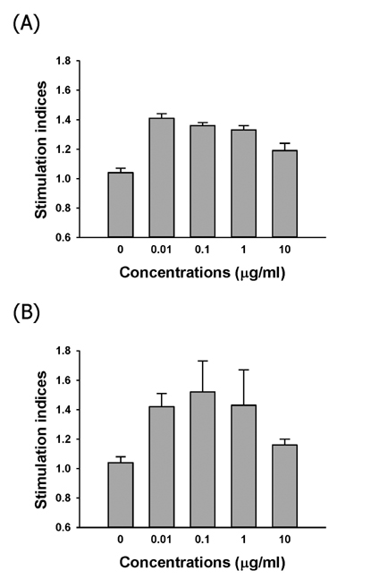
Figure 1 Level of splenocyte proliferation induced by various concentrations of in vitro CBG-CS-2 treatment together with (A) T cell mitogen, Con A and (B) B cell mitogen, LPS. Stimulation indices were calculated by dividing the CPM of the sample from the CBG-CSG-2 treatment with that of the control non-treated sample. The CPM of the control non-treated sample was (A) 75,298.67±1997.07 and (B) 8,857.67±242.61.
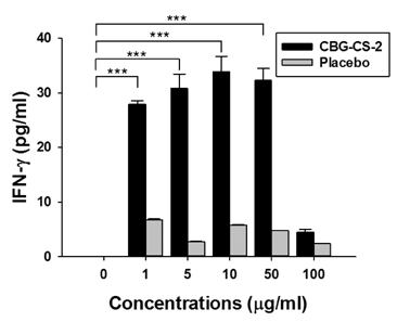
Figure 2 Stimulation of Th1-type cytokine expression by in vitro stimulation of splenocytes with CBG-CS-2. Levels of IFN-γ in the splenocyte culture supernatant after in vitro stimulation with the indicated concentration of CBG-CS-2 or placebo were determined in the presence of 1 µg/ml ConA. ***p<0.001 indicates a significant difference in the results obtained.

Figure 3 Stimulating activity of in vitro treatment of CBG-CS-2 on NK-92MI-mediated cytotoxicity against K562 target cells. (A) NK-92MI cells were stimulated with CBG-CS-2 for 144 hrs and incubated with 51 Cr-labelled K562 target cells with an E:T ratio of 200:1. A similar pattern of cytotoxicity was observed when E:T ratios of 100:1 and 50:1 were used (data not shown). (B) NK-92MI cells were stimulated with either CBG-CS-2 or a placebo for 144 hrs and incubated with 51 Cr-labelled K562 target cells with indicated E:T ratios. ***p<0.001 indicates a significant difference in the results obtained.
Reference
-
1. Sung JM, Lee HK, Choi YS, Kim YY, Kim SH, Sung GH. Distribution and taxonomy of entomopathogenicfungal species from Korea. Korean J Mycol. 1997; 25:239–252.2. Sung JM, Kim CH, Yang KJ, Lee HK, Kim YS. Studies on distribution and utilization of Cordyceps militaris and C. nutans. Korean J Mycol. 1993; 21:94–105.3. Sung JM, Lee HK, Yoo YJ, Choi YS, Kim SH, Kim YO, Sung SH. Classification of Cordyceps species based on protein banding pattern. Korean J Mycol. 1998; 26:1–7.4. Sung GH, Hywel-Jones NL, Sung JM, Luangsa-ard JJ, Shrestha B, Spatafora JW. Phylogenetic classification of Cordyceps and the clavicipitaceous fungi. Stud Mycol. 2007; 57:5–59.
Article5. Jang YS, Hong SW. Notes on unrecorded fresh fungi of Cordyceps in Korea. Korean J Mycol. 1986; 14:85–88.6. Tuli HS, Sandhu SS, Sharma AK. Pharmacological and therapeutic of Cordyceps with special reference to Codycepin. 3 Biotech. 2014; 4:1–12.
Article7. Holliday JC, Cleaver MP. Medicinal value of the caterpillar fungi species of the genus Cordyceps (Fr.) Link (Ascomycetes). A review. Int J Med Mushr. 2008; 10:219–234.
Article8. Wang SY, Shiao MS. Pharmacological functions of Chinese medicinal fungus Cordyceps sinensis and related species. J Food Drug Anal. 2000; 8:248–257.9. Zhou X, Gong Z, Su Y, Lin J, Tang K. Cordyceps fungi: natural products, pharmacological functions and developmental products. J Pharm Pharmacol. 2009; 61:279–291.
Article10. Zhu JS, Halpern GM, Jones K. The scientific rediscovery of an ancient Chinese herbal medicine: Cordyceps sinensis. J Altern Complement Med. 1998; 4:289–303.
Article11. Siu KM, Mak DHF, Chiu PY, Poon MKT, Du Y, Ko KM. Pharmrcological basis of 'Yin-nourishing' and 'Yang-invigorating' actions of Cordyceps, a Chinese tonifying herb. Life Sci. 2004; 76:385–395.
Article12. Khan MA, Tania M, Zhang D, Chen H. Cordyceps mushroom: A potent anticancer nutraceutical. Open Nutraceut J. 2010; 3:179–183.13. Liu WC, Wang SC, Tsai ML, Chen MC, Wang YC, Hong JH, McBride WH, Chiang CS. Protection against radiation-induced bone marrow and intestinal injuries by Cordyceps sinensis, a Chinese herbal medicine. Radiat Res. 2006; 166:900–907.
Article14. Ko WS, Hsu SL, Chyau CC, Chen KC, Peng RY. Compound Cordyceps TCM-700C exhibits potent hepatoprotective capability in animal model. Fitoterapia. 2010; 81:1–7.
Article15. Kiho T, Hui J, Yamane A, Ukai S. Polysaccharides in fungi. XXXII. Hypoglycemic activity and chemical properties of a polysaccharide from the cultural mycelium of Cordyceps sinensis. Biol Pharm Bull. 1993; 16:1291–1293.
Article16. Kiho T, Yamane A, Hui J, Usui S, Ukai S. Polysaccharides in fungi. XXXVI. Hypoglycemic activity of a polysaccharide (CS-F30) from the cultural mycelium of Cordyceps sinensis and its effect on glucose metabolism in mouse liver. Biol Pharm Bull. 1996; 19:294–296.
Article17. Zhao CS, Yin WT, Wang JY, Zhang Y, Yu H, Cooper R, Smidt C, Zhu JS. CordyMax Cs-4 improves glucose metabolism and increases insulin sensitivity in normal rats. J Altern Complement Med. 2002; 8:309–314.
Article18. Lo HC, Tu ST, Lin KC, Lin SC. The anti-hyperglycemic activity of the fruiting body of Cordyceps in diabetic rats induced by nicotinamide and streptozotocin. Life Sci. 2004; 74:2897–2908.
Article19. Li SP, Zhang GH, Zeng Q, Huang ZG, Wang YT, Dong TTX, Tsim KWK. Hypoglycemic activity of polysaccharide, with antioxidation, isolated from cultured Cordyceps mycelia. Phytomedicine. 2006; 13:428–433.
Article20. Kuo CF, Chen CC, Luo YH, Huang RY, Chuang WJ, Sheu CC, Lin YS. Cordyceps sinensis mycelium protects mice from group A streptococcal infection. J Med Microbiol. 2005; 54:795–802.
Article21. Jang SH, Kim SH, Lee HY, Jang SH, Jang H, Chae SW, Jung SJ, So BO, Ha KC, Sin HS, Jang YS. Immune-modulating activity of extract prepared from mycelial culture of Chinese caterpillar mushroom, Ophiocordyceps sinensis (Ascomycetes). Int J Med Mushrooms. 2015; 17:1189–1199.
Article22. Langhans B, Ahrendt M, Nattermann J, Sauerbruch T, Spengler U. Comparative study of NK cell-mediated cytotoxicity using radioactive and flow cytometric cytotoxicity assays. J Immunol Methods. 2005; 306:161–168.
Article
- Full Text Links
-
- Actions
-
Cited
- CITED
-
- Close
- Share
-
- Similar articles
-
- Growth and Cultural Characteristics of Ophiocordyceps longissima Collected in Korea
- Lung Cancer Organoid System to Evaluate the Cytotoxicity of Natural Killer Cells
- Cultural Characteristics of Ophiocordyceps heteropoda Collected from Korea
- Ethanol Extract of Chaenomeles sinensis Inhibits the Development of Benign Prostatic Hyperplasia by Exhibiting Anti-oxidant and Anti-inflammatory Effects
- Diverse Expression of NK Cell Receptor between Fetal Thymocytes and Fetal Liver Lymphocytes from the Same Individuals
