Lactoferrin Stimulates Mouse Macrophage to Express BAFF via Smad3 Pathway
- Affiliations
-
- 1Department of Molecular Bioscience, College of Biomedical Science, Kangwon National University, Chuncheon 200-701, Korea. phkim@kangwon.ac.kr
- 2Division of Animal Life and Environmental Science, College of Agriculture and Life Science, Hankyong National University, Anseong 456-749, Korea.
- KMID: 2168005
- DOI: http://doi.org/10.4110/in.2012.12.3.84
Abstract
- B cell-activating factor belonging to the TNF family (BAFF) is primarily expressed by macrophages and stimulates B cell proliferation, differentiation, survival, and Ig production. In this study, we explored the effect of lactoferrin (LF) on BAFF expression by murine macrophages. We determined the level of BAFF expression at the transcriptional and protein levels using RT-PCR and ELISA, respectively. LF markedly enhanced BAFF expression in mouse macrophages at both the transcriptional and protein levels. Overexpression of Smad3/4 further increased LF-induced BAFF transcription while DN-Smad3 abolished the LF-induced BAFF expression. These results demonstrate that LF can enhance BAFF expression through Smad3/4 pathway.
Keyword
MeSH Terms
Figure
-

Figure 1 LF stimulates mouse macrophages to express BAFF. (A) Effect of LF on BAFF transcriptional levels in peritoneal mouse macrophages. Freshly isolated mouse peritoneal macrophages were incubated with indicated dose of LF for 24 h. BAFF mRNA levels were determined by RT-PCR. (B) Effect of LF on the expression of BAFF at the protein level. Macrophages were treated with LF (60µg/ml) for 48 h and the secreting form of BAFF was measured by ELISA. (C) Effect of LF on BAFF transcriptional level in mouse macrophages. RAW264.7 cells (mouse macrophage cell line) were treated with indicated dose of LF for 24 h (left panel). LF (60µg/ml) was added to the cell cultures for the indicated times (right panel). BAFF mRNA levels were determined by RT-PCR. Fold increase (Fold I.) values represent relative amounts of BAFF cDNA normalized to the expression of β-actin cDNA using Kodak Molecular Imaging software.
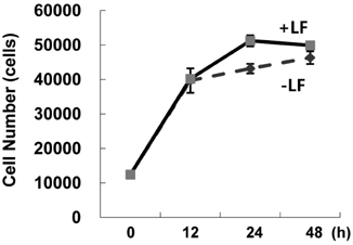
Figure 2 LF has marginal effect on the proliferation of macrophages. RAW264.7 cells were incubated with LF (60µg/ml) for indicated times. Cell proliferation was assessed using a cell counting kit 8. The viable cell number was determined based on OD values measured for standard curve. Data are means of triplicate samples±SEM.
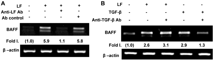
Figure 3 LF in itself stimulates macrophages to express BAFF. (A) LF (60µg/ml) was pre-treated with anti-LF antiserum (diluted to 1:8) for 1 h and added to RAW264.7 cells, incubated for 18 h. Levels of BAFF transcription were determined by RT-PCR. (B) LF (60µg/ml), TGF-β (1 ng/ml) and pan anti-TGF-β Ab (5µg/ml) were added to RAW264.7 cell and incubated for 18 h. Before addition of LF to culture, LF was pre-treated with anti-TGF-β Ab for 1 h. Levels of BAFF transcripts were determined by RT-PCR.
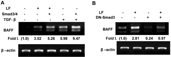
Figure 4 LF enhances BAFF expression through Smad3 pathway. RAW264.7 cells (1×10⁶) were transfected with Smad3/Smad4 (each 1 µg) (A) and DN-Smad3 expression vector (10µg) (B) for 24 h and stimulated with LF (60µg/ml) for 18 h. Levels of BAFF transcripts were determined by RT-PCR.
Cited by 1 articles
-
Oral Administration of Silk Peptide Enhances the Maturation and Cytolytic Activity of Natural Killer Cells
Sun-Hee Jang, Mi-Sun Oh, Hyang-Im Baek, Ki-Chan Ha, Jeong-Yong Lee, Yong-Suk Jang
Immune Netw. 2018;18(5):. doi: 10.4110/in.2018.18.e37.
Reference
-
1. Metz-Boutigue MH, Jollès J, Mazurier J, Schoentgen F, Legrand D, Spik G, Montreuil J, Jollès P. Human lactotransferrin: amino acid sequence and structural comparisons with other transferrins. Eur J Biochem. 1984. 145:659–676.
Article2. Legrand D, Elass E, Carpentier M, Mazurier J. Lactoferrin: a modulator of immune and inflammatory responses. Cell Mol Life Sci. 2005. 62:2549–2559.3. Valenti P, Antonini G. Lactoferrin: an important host defence against microbial and viral attack. Cell Mol Life Sci. 2005. 62:2576–2587.4. Puddu P, Valenti P, Gessani S. Immunomodulatory effects of lactoferrin on antigen presenting cells. Biochimie. 2009. 91:11–18.
Article5. Puddu P, Latorre D, Carollo M, Catizone A, Ricci G, Valenti P, Gessani S. Bovine lactoferrin counteracts Toll-like receptor mediated activation signals in antigen presenting cells. PLoS One. 2011. 6:e22504.
Article6. Craxton A, Magaletti D, Ryan EJ, Clark EA. Macrophage- and dendritic cell--dependent regulation of human B-cell proliferation requires the TNF family ligand BAFF. Blood. 2003. 101:4464–4471.
Article7. Dubois B, Vanbervliet B, Fayette J, Massacrier C, Van Kooten C, Brière F, Banchereau J, Caux C. Dendritic cells enhance growth and differentiation of CD40-activated B lymphocytes. J Exp Med. 1997. 185:941–951.
Article8. Fagarasan S, Honjo T. T-Independent immune response: new aspects of B cell biology. Science. 2000. 290:89–92.
Article9. Groom J, Kalled SL, Cutler AH, Olson C, Woodcock SA, Schneider P, Tschopp J, Cachero TG, Batten M, Wheway J, Mauri D, Cavill D, Gordon TP, Mackay CR, Mackay F. Association of BAFF/BLyS overexpression and altered B cell differentiation with Sjögren's syndrome. J Clin Invest. 2002. 109:59–68.
Article10. Mackay F, Ambrose C. The TNF family members BAFF and APRIL: the growing complexity. Cytokine Growth Factor Rev. 2003. 14:311–324.
Article11. Sutherland AP, Mackay F, Mackay CR. Targeting BAFF: immunomodulation for autoimmune diseases and lymphomas. Pharmacol Ther. 2006. 112:774–786.
Article12. Park SR, Lee JH, Kim PH. Smad3 and Smad4 mediate transforming growth factor-beta1-induced IgA expression in murine B lymphocytes. Eur J Immunol. 2001. 31:1706–1715.
Article13. Imamura T, Takase M, Nishihara A, Oeda E, Hanai J, Kawabata M, Miyazono K. Smad6 inhibits signalling by the TGF-beta superfamily. Nature. 1997. 389:622–626.14. Goto D, Yagi K, Inoue H, Iwamoto I, Kawabata M, Miyazono K, Kato M. A single missense mutant of Smad3 inhibits activation of both Smad2 and Smad3, and has a dominant negative effect on TGF-beta signals. FEBS Lett. 1998. 430:201–204.
Article15. Kim HA, Jeon SH, Seo GY, Park JB, Kim PH. TGF-beta1 and IFN-gamma stimulate mouse macrophages to express BAFF via different signaling pathways. J Leukoc Biol. 2008. 83:1431–1439.
Article16. Zemann N, Klein P, Wetzel E, Huettinger F, Huettinger M. Lactoferrin induces growth arrest and nuclear accumulation of Smad-2 in HeLa cells. Biochimie. 2010. 92:880–884.
Article
- Full Text Links
-
- Actions
-
Cited
- CITED
-
- Close
- Share
-
- Similar articles
-
- Activin A Stimulates Mouse APCs to Express BAFF via ALK4-Smad3 Pathway
- B-cell-activating factor is a regulator of adipokines and a possible mediator between adipocytes and macrophages
- Effect of Bovine and Human Lactoferrin on MA 104 Cell Infected with Human Rotavirus
- Serum and Cerebrospinal Fluid(CSF) Nitric Oxide, Macrophage Inflammatory Protein-1 alpha and Lactoferrin Levels in Aseptic Meningitis
- TGF-beta1 induces mouse dendritic cells to express VEGF and its receptor (Flt-1) under hypoxic conditions
