The Healing Effects of Autologous Mucosal Grafts in Experimentally Injured Rabbit Maxillary Sinuses
- Affiliations
-
- 1Department of Otorhinolaryngology, Kocaeli University Faculty of Medicine, Kocaeli, Turkey. doktor.kbb@hotmail.com
- 2Department of Histology and Embryology, Akdeniz University Faculty of Medicine, Antalya, Turkey.
- 3Department of Otorhinolaryngology, Acıbadem University Faculty of Medicine, Istanbul, Turkey.
- KMID: 2166286
- DOI: http://doi.org/10.21053/ceo.2016.9.1.44
Abstract
OBJECTIVES
Healing processes of the nose and paranasal sinuses are quite complex, and poorly understood. In this study, we aimed to compare the effect of mucosal autologous grafts on the degenerated rabbit maxillary sinus mucosa with spontaneous wound healing. It is hypothesized that mucosal grafts will enhance ciliogenesis and improve the morphology of regenerated cilia.
METHODS
Ten female New Zealand rabbits were included in the study. They underwent external maxillary sinus surgery through a transcutaneous approach. A total of 20 maxillary sinuses were randomly divided into 2 groups: 'spontaneous healing group' and 'autologous graft group.' The animals were sacrificed at the 14th day after the surgery. Scanning electron microscope (SEM), and light microscope were used for the evaluation.
RESULTS
Cellular composition of the graft group is better than the spontaneous healing group. The graft group had larger areas covered with ciliary epithelium than the spontaneous healing group, and the mean length of the cilias were also longer. Additionally, there were wider cilia with abnormal morphology areas in the spontaneous healing group.
CONCLUSION
In our opinion, covering of the denuded areas with a graft improves re-epithelization, and may prevent the early complications after sinus surgeries.
Keyword
MeSH Terms
Figure
-
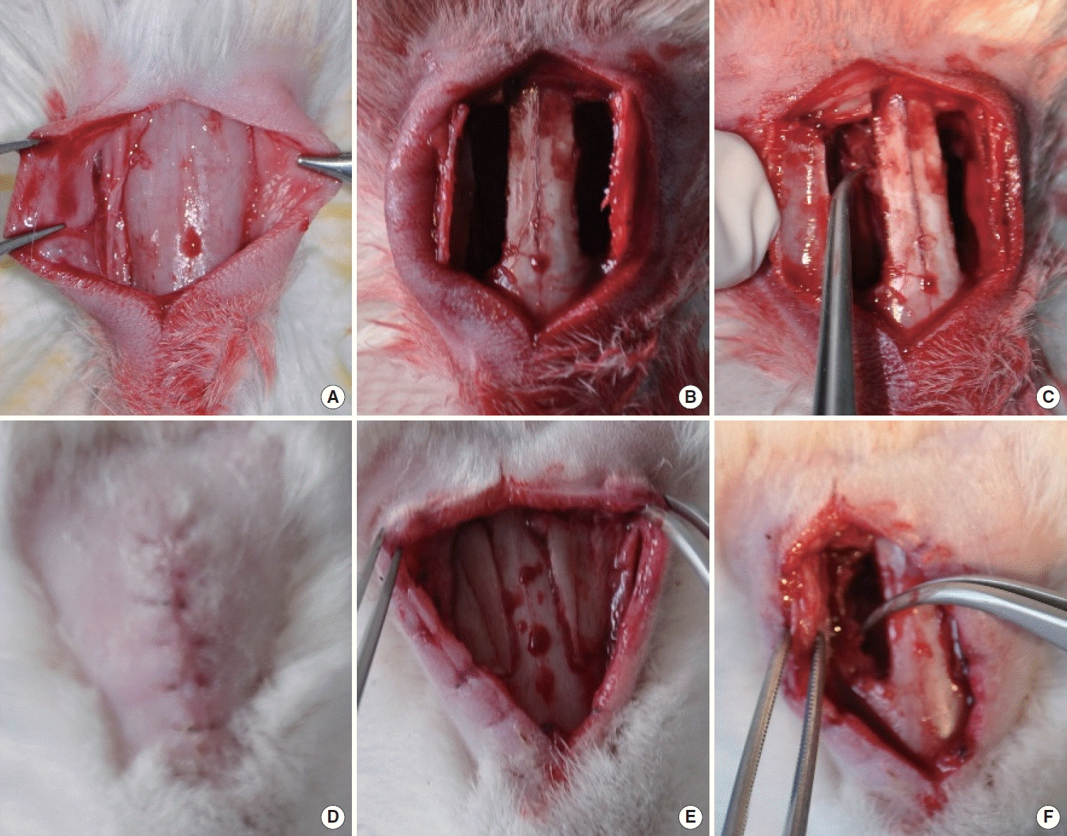
Fig. 1. Anterior walls of the maxillary sinuses (A), bilaterally bone flaps (B), elevation of an approximately 1-cm2 mucosal area on the medial maxillary wall (C), healed animal (D), healed anterior wall of the sinus (E), inflamed and hypertrophic mucosa with neo osteogenesis (F).
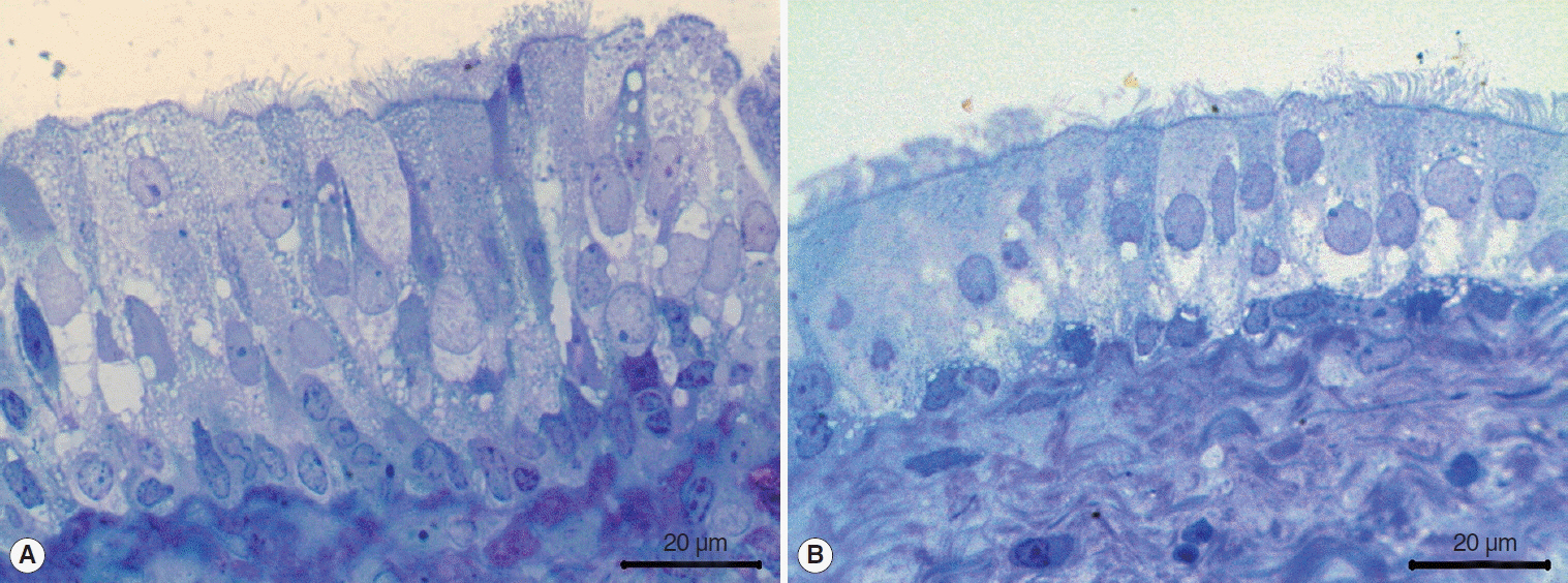
Fig. 2. Light microscopic examinations (toluidin blue) showed graft group (A) contained more and higher epithelial cells than spontaneous healing group (B).
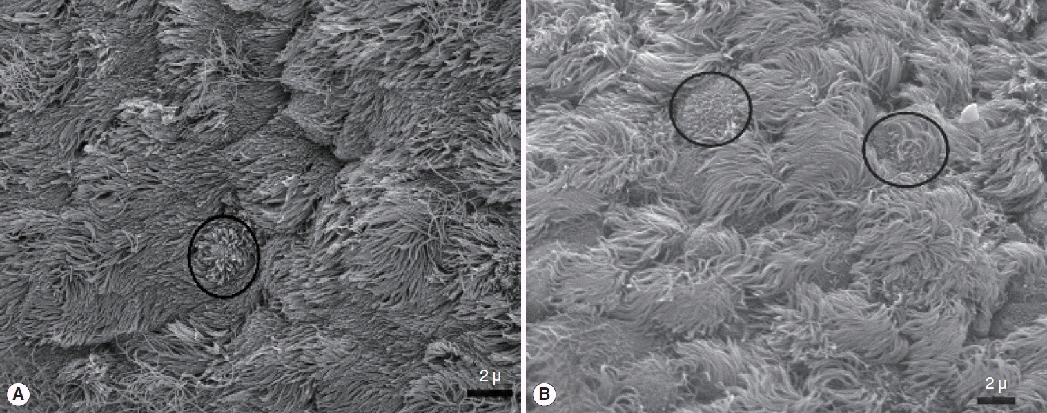
Fig. 3. Scanning electron microscope images of epithelial ciliary organizations. Graft (A) and spontaneous healing group (B). Apical surface buds are seen early of the epitelial ciliary development both of the graft and spontaneous healing groups. They are showed with ring in the A and B panels.
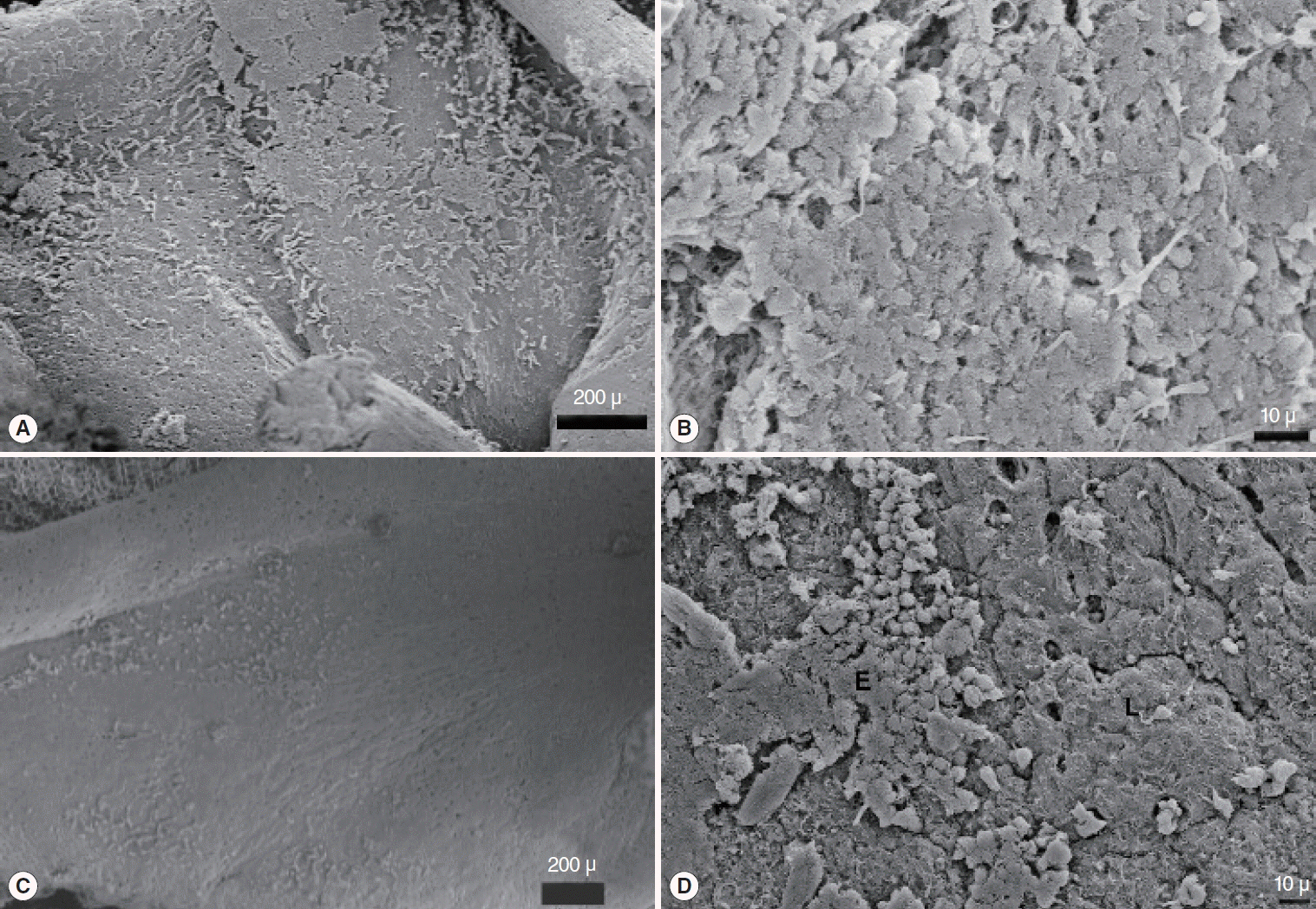
Fig. 4. Cell cordons in the graft group were spreaded over the surface to form a superficial cover; graft (A, B), epithelial dispersion was in different areas and unable to create a cover; spontaneous healing group (C, D).

Fig. 5. Abnormal ciliary formations in the scanning electron microscope images. Graft (A) and spontaneous healing group (B).
Reference
-
1. Thawley SE, Deddens AE. Transfrontal endoscopic management of frontal recess disease. Am J Rhinol. 1995; Nov-Dec. 9(6):307–11.
Article2. Wormald PJ. The axillary flap approach to the frontal recess. Laryngoscope. 2002; Mar. 112(3):494–9.
Article3. Wormald PJ, Chan SZ. Surgical techniques for the removal of frontal recess cells obstructing the frontal ostium. Am J Rhinol. 2003; Jul-Aug. 17(4):221–6.
Article4. Watelet JB, Bachert C, Gevaert P, Van Cauwenberge P. Wound healing of the nasal and paranasal mucosa: a review. Am J Rhinol. 2002; Mar-Apr. 16(2):77–84.
Article5. Casteleyn C, Cornillie P, Hermens A, Van Loo D, Van Hoorebeke L, van den Broeck W, et al. Topography of the rabbit paranasal sinuses as a prerequisite to model human sinusitis. Rhinology. 2010; Sep. 48(3):300–4.
Article6. Koybasioglu A, Ileri F, Beder L, Inal E. Anatomy of the rabbit maxillary sinus. Kulak Burun Bogaz Bas Boyun Cerrahi Derg. 1997; 5:41–4.7. Inanli S, Tutkun A, Batman C, Okar I, Uneri C, Sehitoglu MA. The effect of endoscopic sinus surgery on mucociliary activity and healing of maxillary sinus mucosa. Rhinology. 2000; Sep. 38(3):120–3.8. Bassiouny A, Atef AM, Raouf MA, Nasr SM, Nasr M, Ayad EE. Ultrastructural ciliary changes of maxillary sinus mucosa following functional endoscopic sinus surgery: an image analysis quantitative study. J Laryngol Otol. 2003; Apr. 117(4):273–9.
Article9. Guo Y, Majima Y, Hattori M, Seki S, Sakakura Y. Effects of functional endoscopic sinus surgery on maxillary sinus mucosa. Arch Otolaryngol Head Neck Surg. 1997; Oct. 123(10):1097–100.
Article10. Perez AC, Cunha Junior Ada S, Fialho SL, Silva LM, Dorgam JV, Murashima Ade A, et al. Assessing the maxillary sinus mucosa of rabbits in the presence of biodegradable implants. Braz J Otorhinolaryngol. 2012; Dec. 78(6):40–6.
Article11. Swibel Rosenthal LH, Benninger MS, Stone CH, Zacharek MA. Wound healing in the rabbit paranasal sinuses after Coblation: evaluation for use in endoscopic sinus surgery. Am J Rhinol Allergy. 2009; May-Jun. 23(3):360–3.12. Forsgren K, Stierna P, Kumlien J, Carlsoo B. Regeneration of maxillary sinus mucosa following surgical removal. Experimental study in rabbits. Ann Otol Rhinol Laryngol. 1993; Jun. 102(6):459–66.13. Benninger MS, Schmidt JL, Crissman JD, Gottlieb C. Mucociliary function following sinus mucosal regeneration. Otolaryngol Head Neck Surg. 1991; Nov. 105(5):641–8.
Article14. Maccabee MS, Trune DR, Hwang PH. Paranasal sinus mucosal regeneration: the effect of topical retinoic acid. Am J Rhinol. 2003; May-Jun. 17(3):133–7.
Article15. Hwang PH, Chan JM. Retinoic acid improves ciliogenesis after surgery of the maxillary sinus in rabbits. Laryngoscope. 2006; Jul. 116(7):1080–5.
Article16. Beule AG, Scharf C, Biebler KE, Gopferich A, Steinmeier E, Wolf E, et al. Effects of topically applied dexamethasone on mucosal wound healing using a drug-releasing stent. Laryngoscope. 2008; Nov. 118(11):2073–7.
Article17. Beule AG, Steinmeier E, Kaftan H, Biebler KE, Gopferich A, Wolf E, et al. Effects of a dexamethasone-releasing stent on osteoneogenesis in a rabbit model. Am J Rhinol Allergy. 2009; Jul-Aug. 23(4):433–6.
Article18. Hosemann W, Goede U, Sauer M. Wound healing of mucosal autografts for frontal cerebrospinal fluid leaks: clinical and experimental investigations. Rhinology. 1999; Sep. 37(3):108–12.19. Hildenbrand T, Wormald PJ, Weber RK. Endoscopic frontal sinus drainage Draf type III with mucosal transplants. Am J Rhinol Allergy. 2012; Mar-Apr. 26(2):148–51.
Article
- Full Text Links
-
- Actions
-
Cited
- CITED
-
- Close
- Share
-
- Similar articles
-
- Effects of Preservation of the Lamina Propria of the Maxillary Sinus Mucosa on Mucosal Regeneration in Experimentally Induced Maxillary Sinusitis with Polyposis in Rabbits
- Effects of Pseudomonas Aeruginosa Infection and Mechanical Trauma to the Sinus Mucosa on Polyp Formation in the Rabbit Maxillary Sinuses
- A Morphological Study of the Paranasal Sinuses in Koreans
- The Effect of Platelet-Rich Plasma(PRP) on the Survival of the Autologous Fat Graft
- Anatomical variants of paranasal sinus affecting the ostiomeatal unit
