A Case of Nasal Cavity Foreign Bodies by Inhalation of Polyurethane Foam
- Affiliations
-
- 1Department of Otorhinolaryngology-Head and Neck Surgery, Kangbuk Samsung Hospital, Sungkyunkwan University School of Medicine, Seoul, Korea.
- 2Department of Otorhinolaryngology-Head and Neck surgery, Dongtan Sacred Heart Hospital, Hallym University School of Medicine, Hwaseong, Korea. enthsj@hanmail.net
- KMID: 2165158
- DOI: http://doi.org/10.18787/jr.2016.23.1.65
Abstract
- Foreign bodies in the nasal cavity are commonly encountered in otorhinolaryngologic practice, particularly among children and mentally handicapped patients. Such foreign bodies include plastic toys, pebbles, seeds, buttons, and many others. Many of these foreign bodies can be easily removed with simple tools. However, some of them adhere to the nasal mucosa, resulting in complications such as necrosis or neurovascular injury of the nasal mucosa. Polyurethane foam in the nasal cavity has never been reported in Korea. Furthermore, the complications caused by polyurethane foam in the nasal cavity have not yet been reported. In this article, we report a man who presented with polyurethane foam that had spread into both nasal cavity and nasopharynx by inhalation and adhered to the nasal cavity and sinuses.
Keyword
MeSH Terms
Figure
-

Fig. 1. Preoperative endoscopic findings. Whitish foreign bodies surrounding middle turbinate in right nasal cavity (A), inferior turbinate in right nasal cavity (B), middle turbinate in left nasal cavity (C), and in the nasopharynx (D).
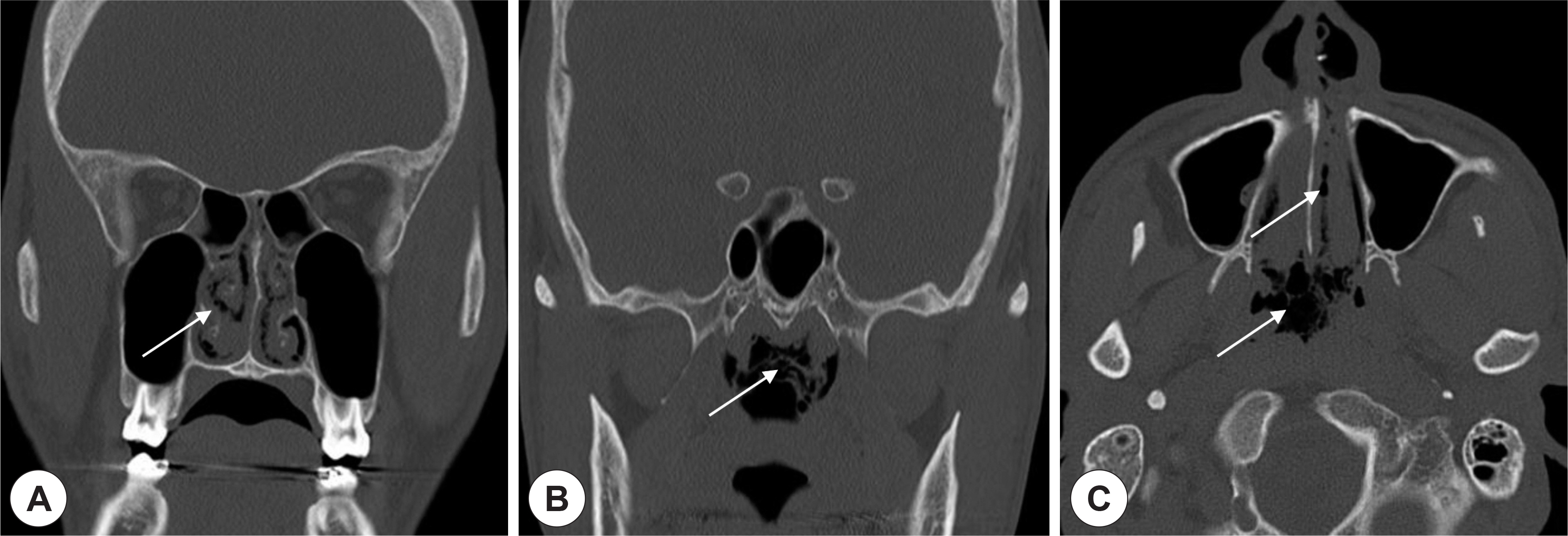
Fig. 2. PNS CT with noncontrast. Soft tissue densities with air bubbles sign (white arrow) were observed in the nasal cavity and the nasopharynx which were shown at coronal view (A), (B) and axial view (C).

Fig. 3. Removed foreign bodies during operation. Multiple pieces of whitish foreign bodies were removed from both nasal cavity and the nasopharynx. Red-colored nasal mucosa attached to foreign bodies.
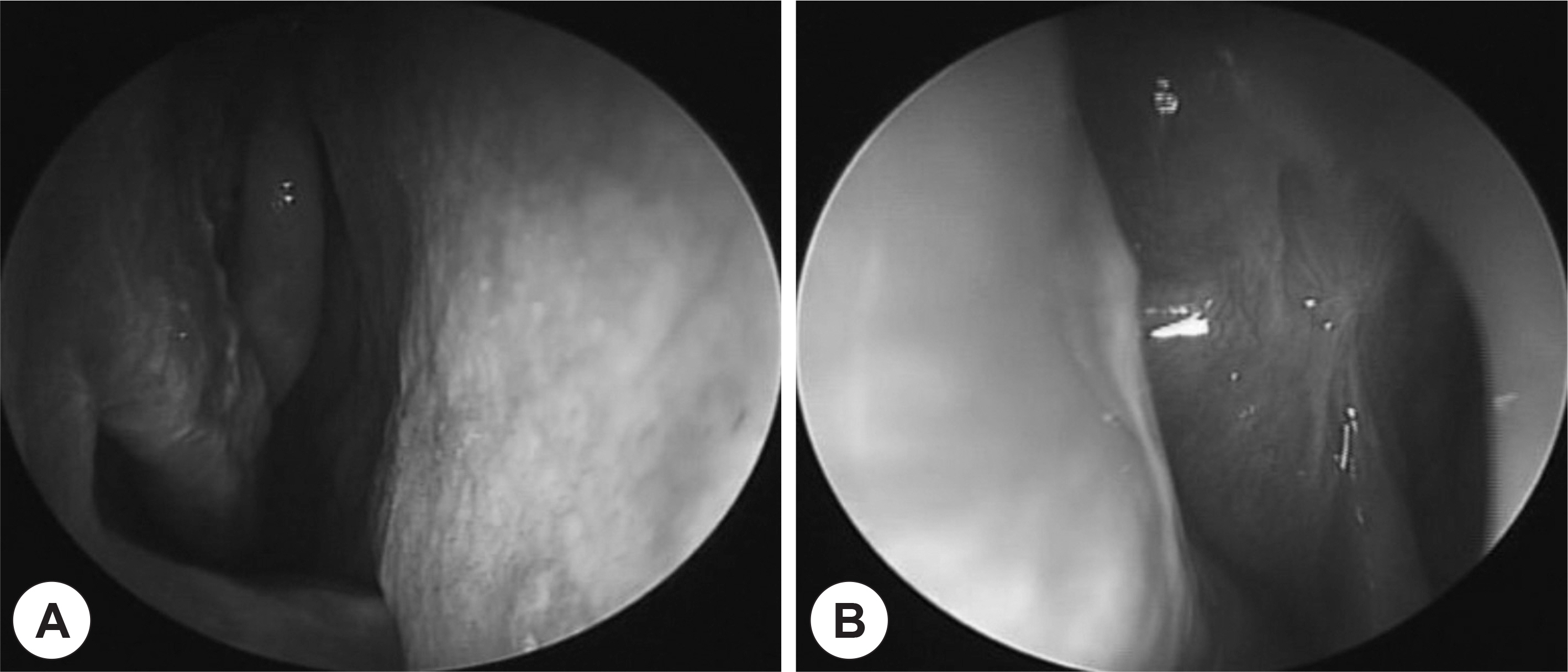
Fig. 4. Postoperative follow-up endoscopic findings (POD 120 days). There were no whitish foreign bodies and no signs of necrosis and inflammation in both nasal cavity. (A) Right nasal cavity (B) Left middle meatus.
Reference
-
References
1). Kalan A, Tariq M. Foreign bodies in the nasal cavities: a comprehensive review of the aetiology, diagnostic pointers, and therapeutic measures. Postgrad Med J. 2000; 76(898):484–7.
Article2). Lee HM, Kim DH, Kim JM, Hwang SJ, Lee SH. Nasal Septal Perforation due to Button Battery. J Rhinol. 2001; 8(1, 2):69–72.3). Figueiredo RR, Azevedo AA, Kos AO, Tomita S. Nasal foreign bodies: description of types and complications in 420 cases. Braz J Otorhinolaryngol. 2006; 72(1):18–23.
Article4). Mangussi-Gomes J, Andrade JS, Matos RC, Kosugi EM, Penido Nde O. ENT foreign bodies: profile of the cases seen at a tertiary hospital emergency care unit. Braz J Otorhinolaryngol. 2013; 79(6):699–703.
Article5). Chiun KC, Tang IP, Tan TY, Jong DE. Review of ear, nose and throat foreign bodies in Sarawak General Hospital. A five year experience. Med J Malaysia. 2012; 67(1):17–20.6). Francois M, Hamrioui R, Narcy P. Nasal foreign bodies in children. Eur Arch Otorhinolaryngol. 1998; 255(3):132–4.7). Claudet I, Salanne S, Debuisson C, Marechal C, Rekhroukh H, Grou-teau E. [Nasal foreign body in infants]. Arch Pediatr. 2009; 16(9):1245–51.8). Ogunleye AO, Sogebi OA. Nasal foreign bodies in the African children. Afr J Med Med Sci. 2004; 33(3):225–8.9). Lee SU, Park MC, Shin HC, Jin SM. A Case of Parapharyngeal Foreign Bodies Causing Explosion of Polyurethane Foam in Oral Cavity. Korean J Otorhinolaryngol-Head Neck Surg. 2011; 54(11):788–90.
Article10). Henriks-Eckerman ML, Valimaa J, Rosenberg C, Peltonen K, Engstrom K. Exposure to airborne isocyanates and other thermal degradation products at polyurethane-processing workplaces. J Environ Monit. 2002; 4(5):717–21.
Article11). Sowerby RJ, Sowerby LJ, Vinden C. A sticky situation: management of spray polyurethane foam insulation in body orifices. CJEM. 2011; 13(6):404–8.12). Dernehl CU. Health hazards associated with polyurethane foams. J Occup Med. 1966; 8(2):59–62.13). Hueper WC. Cancer Induction by Polyurethan and Polysilicone Plas-tics. J Natl Cancer Inst. 1964; 33:1005–27.14). Tsuang W, Huang YC. Asthma induced by exposure to spray polyurethane foam insulation in a residential home. J Occup Environ Med. 2012; 54(3):272–3.
Article
- Full Text Links
-
- Actions
-
Cited
- CITED
-
- Close
- Share
-
- Similar articles
-
- A Case of Parapharyngeal Foreign Bodies Causing Explosion of Polyurethane Foam in Oral Cavity
- Impacted Foreign Body in the Anterior Nasal Cavity Presenting With Tooth Pain
- A Case of Maxillary Sinus Spindle Cell Sarcoma: Alert to Polyurethane Associated Malignancy
- A Case of Septal Perforation Reconstructed with Alloderm Interposition Graft Resulted from Magnetic Nasal Foreign Bodies
- A Case of Metallic Foreign Body Retained in the Naso-Maxillo-Ethmoido-Orbital Complex
