J Rhinol.
2023 Mar;30(1):45-47. 10.18787/jr.2023.00007.
Impacted Foreign Body in the Anterior Nasal Cavity Presenting With Tooth Pain
- Affiliations
-
- 1Department of Otorhinolaryngology-Head and Neck Surgery, Chung-Ang University College of Medicine, Seoul, Republic of Korea
- KMID: 2540858
- DOI: http://doi.org/10.18787/jr.2023.00007
Abstract
- Foreign bodies pose a diagnostic challenge to clinicians, and nasal foreign bodies have the potential to lead to significant morbidity. Although foreign bodies in the nasal cavity are a commonly encountered problem in pediatric patients, a foreign body in the nasal cavity not associated with a trauma history is rare in adults. We recently experienced a 35-year-old man who presented with a foreign body in his right nasal cavity and anterior tooth pain. He was not sure what the material was, and we were not able to confirm the material type preoperatively. However, we found that a very large and thick material was impacted and totally obstructed the right anterior nasal cavity. We surgically removed it as a bone block and confirmed postoperatively that the material was glass. This case provided several lessons, and we would like to share our experience.
Keyword
Figure
-
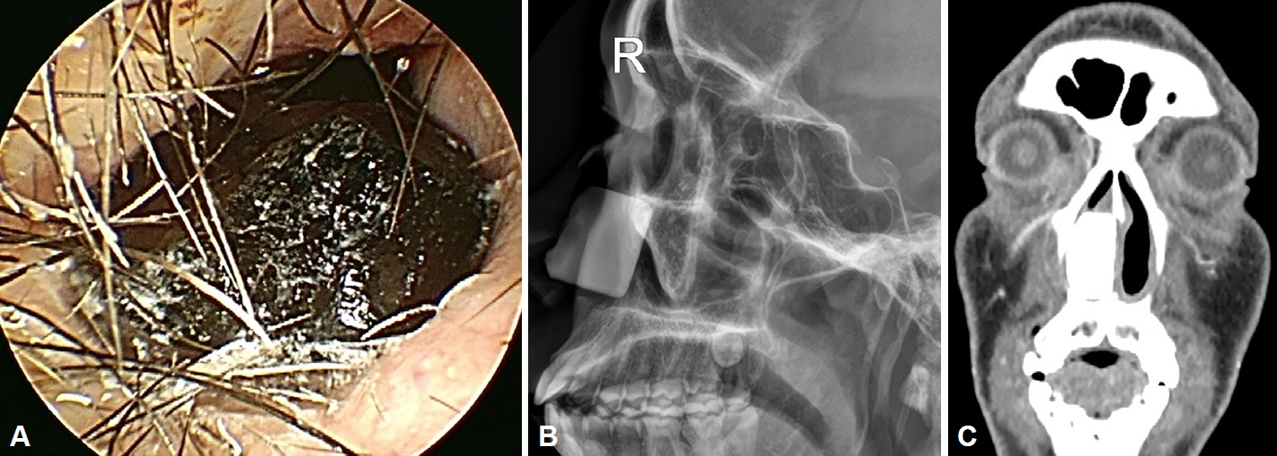
Fig. 1. Endoscopic and imaging findings of the foreign body before surgery. A: Nasal endoscopy showing a dark-colored hard material that totally obstructed the patient’s right nasal cavity. B: Lateral paranasal sinus (PNS) X-ray demonstrating a foreign body measuring approximately 2.5 cm×2.3 cm in the right anterior nasal cavity. C: Coronal PNS computed tomography demonstrating high-density foreign body measuring 2.7 cm×2.2 cm×1.6 cm.

Fig. 2. Glass foreign body removed from the patient’s anterior nasal cavity.
Reference
-
References
1. Yasny JS, Stewart S. Nasal foreign body: an unexpected discovery. Anesth Prog. 2011; 58(3):121–3.
Article2. Ayotunde O, Burkard DJ, Kolacki C, Zamarripa A, Ouellette L, Hamilton M, et al. Nasal foreign body removal: success rates for techniques and devices. Am J Emerg Med. 2022; 56:384–5.
Article3. Yan S, Zeng N, Chen G, Chen Y, Wu Z, Pan H, et al. Presentation and management of nasal foreign bodies in a Chinese metro area. Medicine (Baltimore). 2021; 100(16):e25626.
Article4. Sajid T, Shah MI, Qamar Naqvi SR. Pattern of presentation of nasal foreign bodies, an experience with 155 patients. J Ayub Med Coll Abbottabad. 2018; 30(4):548–50.5. Shueb SS, Boyer HC, Nixdorf DR. Nonodontogenic “tooth pain” of nose and sinus origin. J Am Dent Assoc. 2016; 147(6):457–9.
Article6. Kumar S, Rastogi S, Kumar S, Mahendra P, Bansal M, Chandra L. Pain in trigeminal neuralgia: neurophysiology and measurement: a comprehensive review. J Med Life. 2013; 6(4):383–8.7. Valizadeh S, Pouraliakbar H, Kiani L, Safi Y, Alibakhshi L. Evaluation of visibility of foreign bodies in the maxillofacial region: comparison of computed tomography, cone beam computed tomography, ultrasound and magnetic resonance imaging. Iran J Radiol. 2016; 13(4):e37265.
Article
- Full Text Links
-
- Actions
-
Cited
- CITED
-
- Close
- Share
-
- Similar articles
-
- A Case of Septal Perforation Reconstructed with Alloderm Interposition Graft Resulted from Magnetic Nasal Foreign Bodies
- A Case of a Pharyngeal Impacted Fish Bone Foreign Body Detected by Finger Palpation
- A Case of Metallic Foreign Body Retained in the Naso-Maxillo-Ethmoido-Orbital Complex
- Nasal Septal Perforation due to Button Battery
- Two Cases of Button Battery in Nasal Cavity
