Assessment of anterior-posterior jaw relationships in Korean adults using the nasion true vertical plane in cone-beam computed tomography images
- Affiliations
-
- 1Department of Oral and Maxillofacial Surgery, Hallym University Kangnam Sacred Heart Hospital, Seoul, Korea.
- 2Department of Orthodontics, Hallym University Kangnam Sacred Heart Hospital, Seoul, Korea. ajh0225@hallym.or.kr
- 3Graduate School of Orthodontics, School of Dentistry, University of Nevada Las Vegas, Las Vegas, NV, USA.
- KMID: 2164260
- DOI: http://doi.org/10.4041/kjod.2016.46.3.163
Abstract
OBJECTIVE
The aims of this study were to investigate a simple method for assessing anterior-posterior jaw relationships via cone-beam computed tomography (CBCT) images taken in the natural head position (NHP) relative to the nasion true vertical plane (NTVP), and measure normative data in Korean adults with normal profiles.
METHODS
Subjects were selected from patients presenting for third molar extraction and evaluated as having normal profiles by three examiners. The CBCT images of 80 subjects (39 males, 41 females) were taken in the NHP according to Solow and Tallgren's method. Linear measurements of the A-point, B-point, and Pog were calculated relative to the NTVP. Student's t-test was used to assess sexual differences in these measurements.
RESULTS
The mean linear measurements of the A-point, B-point, and Pog relative to the NTVP were 0.18 mm (standard deviation [SD], 4.77 mm), -4.00 mm (SD, 6.62 mm), and -2.49 mm (SD, 7.14 mm) respectively in Korean males, and 1.48 mm (SD, 4.21 mm), -4.07 mm (SD, 6.70 mm) and -2.91 mm (SD, 7.25 mm) in Korean females respectively. There were no statistically significant differences between Korean males and females (p < 0.05).
CONCLUSIONS
Three-dimensional CBCT analysis using the NTVP is a simple and reliable method for assessing anterior-posterior skeletal relationships.
Keyword
Figure
-
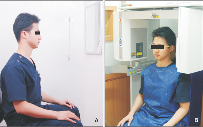
Figure 1 Facial photographs (A) and cone-beam computed tomography images (B) were taken in a sitting position in accordance with Solow and Tallgren's18 method. Patients wore eyeglasses with two fluid levelers attached to the frame.
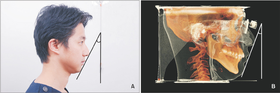
Figure 2 The angle between Ricketts' E-line and the true vertical line was measured in a facial profile photograph (A) and a cone-beam computed tomography image (B).
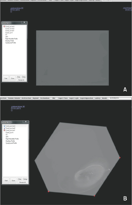
Figure 3 The process of constructing the nasion true vertical plane using In Vivo Dental software. A, A square box was visualized on the volume rendering screen with high brightness. B, Three points were digitized on the bottom surface parallel to the floor.
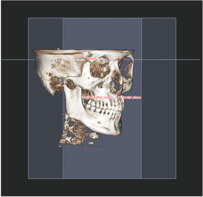
Figure 4 The true horizontal plane, midsagittal plane, and nasion true vertical plane were constructed.

Figure 5 After tracing all lines and digitizing the landmarks required, linear measurements of the A-point, B-point, and Pog relative to the nasion true vertical plane were calculated automatically via In Vivo Dental software (version 5.3; Anatomage Co., San Jose, CA, USA).
Reference
-
1. Sassouni V. A classification of skeletal facial types. Am J Orthod. 1969; 55:109–123.
Article2. Downs WB. Variations in facial relationships; their significance in treatment and prognosis. Am J Orthod. 1948; 34:812–840.
Article3. Steiner CC. Cephalometrics for you and me. Am J Orthod. 1953; 39:729–755.
Article4. McNamara JA Jr. A method of cephalometric evaluation. Am J Orthod. 1984; 86:449–469.
Article5. Lundström F, Lundström A. Natural head position as a basis for cephalometric analysis. Am J Orthod Dentofacial Orthop. 1992; 101:244–247.
Article6. Bjerin R. A comparison between the Frankfort horizontal and the sella turcica-nasion line as reference planes in cephalometric analysis. Acta Odontol Scand. 1957; 15:1–12.7. Moorrees CFA, Kean MR. Natural head position: A basic consideration in the interpretation of cephalometric radiographs. Am J Phys Anthropol. 1958; 16:213–234.
Article8. Lundström A, Lundström F, Lebret LM, Moorrees CF. Natural head position and natural head orientation: basic considerations in cephalometric analysis and research. Eur J Orthod. 1995; 17:111–120.
Article9. Cooke MS, Wei SH. The reproducibility of natural head posture: a methodological study. Am J Orthod Dentofacial Orthop. 1988; 93:280–288.
Article10. Michiels LY, Tourne LP. Nasion true vertical: a proposed method for testing the clinical validity of cephalometric measurements applied to a new cephalometric reference line. Int J Adult Orthodon Orthognath Surg. 1990; 5:43–52.11. Leitão P, Nanda RS. Relationship of natural head position to craniofacial morphology. Am J Orthod Dentofacial Orthop. 2000; 117:406–417.
Article12. Yamamoto K, Ueno K, Seo K, Shinohara D. Development of dento-maxillofacial cone beam X-ray computed tomography system. Orthod Craniofac Res. 2003; 6:Suppl 1. 160–162.
Article13. Nalçaci R, Oztürk F, Sökücü O. A comparison of two-dimensional radiography and threedimensional computed tomography in angular cephalometric measurements. Dentomaxillofac Radiol. 2010; 39:100–106.
Article14. Swennen GR, Schutyser F, Barth EL, De Groeve P, De Mey A. A new method of 3-D cephalometry Part I: the anatomic Cartesian 3-D reference system. J Craniofac Surg. 2006; 17:314–325.15. Cavalcanti MG, Vannier MW. Quantitative analysis of spiral computed tomography for craniofacial clinical applications. Dentomaxillofac Radiol. 1998; 27:344–350.
Article16. Kawamata A, Ariji Y, Langlais RP. Three-dimensional computed tomography imaging in dentistry. Dent Clin North Am. 2000; 44:395–410.17. Cochrane SM, Cunningham SJ, Hunt NP. A comparison of the perception of facial profile by the general public and 3 groups of clinicians. Int J Adult Orthodon Orthognath Surg. 1999; 14:291–295.18. Solow B, Tallgren A. Natural head position in standing subjects. Acta Odontol Scand. 1971; 29:591–607.
Article19. Dahlberg G. Statistical methods for medical and biological students. New York: Interscience Publications;1940.20. Madsen DP, Sampson WJ, Townsend GC. Craniofacial reference plane variation and natural head position. Eur J Orthod. 2008; 30:532–540.
Article21. Weber DW, Fallis DW, Packer MD. Three-dimensional reproducibility of natural head position. Am J Orthod Dentofacial Orthop. 2013; 143:738–744.
Article22. Arnett GW, Jelic JS, Kim J, Cummings DR, Beress A, Worley CM Jr, et al. Soft tissue cephalometric analysis: diagnosis and treatment planning of dentofacial deformity. Am J Orthod Dentofacial Orthop. 1999; 116:239–253.23. Trpkova B, Major P, Prasad N, Nebbe B. Cephalometric landmarks identification and reproducibility: a meta analysis. Am J Orthod Dentofacial Orthop. 1997; 112:165–170.
Article24. Damstra J, Fourie Z, Ren Y. Simple technique to achieve a natural position of the head for cone beam computed tomography. Br J Oral Maxillofac Surg. 2010; 48:236–238.
Article25. Soncul M, Bamber MA. The reproducibility of the head position for a laser scan using a novel morphometric analysis for orthognathic surgery. Int J Oral Maxillofac Surg. 2000; 29:86–90.
Article26. Xia JJ, McGrory JK, Gateno J, Teichgraeber JF, Dawson BC, Kennedy KA, et al. A new method to orient 3-dimensional computed tomography models to the natural head position: a clinical feasibility study. J Oral Maxillofac Surg. 2011; 69:584–591.
Article27. Walker F, Ayoub AF, Moos KF, Barbenel J. Face bow and articulator for planning orthognathic surgery: 1 face bow. Br J Oral Maxillofac Surg. 2008; 46:567–572.
Article28. Hsung TC, Lo J, Li TS, Cheung LK. Recording of natural head position using stereophotogrammetry: a new technique and reliability study. J Oral Maxillofac Surg. 2014; 72:2256–2261.
Article29. Uşümez S, Orhan M. Reproducibility of natural head position measured with an inclinometer. Am J Orthod Dentofacial Orthop. 2003; 123:451–454.
Article30. Brodal A, Pompeniano O. Basic aspects of central vestibular mechanisms. Amsterdam, Netherlands: Elsevier;1972.31. Havard Editorial Board. How to soothe a sore neck. The essentials are icing and heat, gentle therapeutic exercise, and good posture. Harv Mens Health Watch. 2014; 18:5.
- Full Text Links
-
- Actions
-
Cited
- CITED
-
- Close
- Share
-
- Similar articles
-
- Evaluation of buccolingual molar inclinations among different vertical facial types
- Pharyngeal airway analysis of different craniofacial morphology using cone-beam computed tomography (CBCT)
- Use of an anatomical mid-sagittal plane for 3-dimensional cephalometry: A preliminary study
- Stafne bone cavity and cone-beam computed tomography: a report of two cases
- Comparison of three midsagittal planes for three-dimensional cone beam computed tomography head reorientation
