Use of an anatomical mid-sagittal plane for 3-dimensional cephalometry: A preliminary study
- Affiliations
-
- 1Department of Oral and Maxillofacial Sciences, School of Dentistry, Sapienza University of Rome, Rome, Italy. roberto.vernucci@uniroma1.it
- 2Department of Public Health Infectious Diseases, Sapienza University of Rome, Rome, Italy.
- KMID: 2450182
- DOI: http://doi.org/10.5624/isd.2019.49.2.159
Abstract
- PURPOSE
Cone-beam computed tomography (CBCT) is widely used for 3-dimensional assessments of cranio-maxillofacial relationships, especially in patients undergoing orthognathic surgery. We have introduced, for reference in CBCT cephalometry, an anatomical mid-sagittal plane (MSP) identified by the nasion, the midpoint between the posterior clinoid processes of the sella turcica, and the basion. The MSP is an updated version of the median plane previously used at our institution for 2D posterior-anterior cephalometry. This study was conducted to test the accuracy of the CBCT measures compared to those obtained using standard posterior-anterior cephalometry.
MATERIALS AND METHODS
Two operators measured the inter-zygomatic distance on 15 CBCT scans using the MSP as a reference plane, and the CBCT measurements were compared with measurements made on patients' posterior-anterior cephalograms. The statistical analysis evaluated the absolute and percentage differences between the 3D and 2D measurements.
RESULTS
As demonstrated by the absolute mean difference (roughly 1 mm) and the percentage difference (less than 3%), the MSP showed good accuracy on CBCT compared to the 2D plane, especially for measurements of the left side. However, the CBCT measurements showed a high standard deviation, indicating major variability and low precision.
CONCLUSION
The anatomical MSP can be used as a reliable reference plane for transverse measurements in 3D cephalometry in cases of symmetrical or asymmetrical malocclusion. In patients who suffer from distortions of the skull base, the identification of landmarks might be difficult and the MSP could be unreliable. Becoming familiar with the relevant software could reduce errors and improve reliability.
MeSH Terms
Figure
-
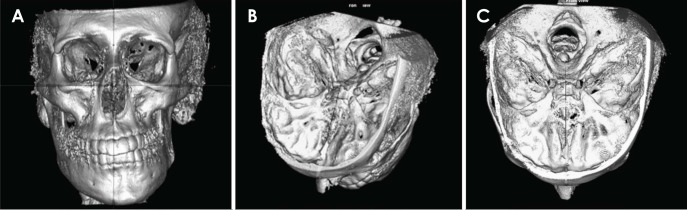
Fig. 1 Landmarks selected for tracing the mid-sagittal plane on cone-beam computed tomographic (CBCT) images. A. Localisation of the nasion. B. Localisation of the midpoint between the posterior clinoid processes. C. Localisation of the basion.
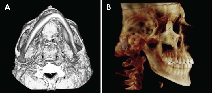
Fig. 2 The anatomical mid-sagittal plane traced by the software on a patient's cone-beam computed tomography 3-dimensional reconstruction. A. Axial orientation. B. Sagittal orientation.
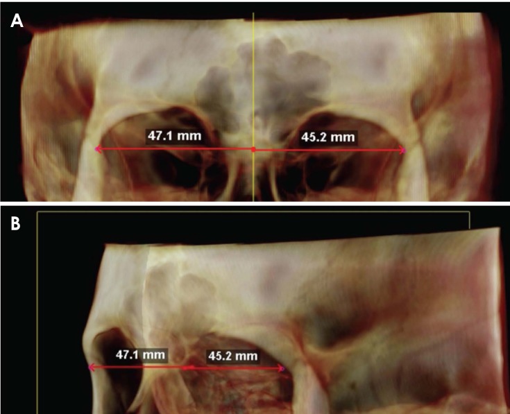
Fig. 3 Different views of the “Dist. to midline” function used to measure the distance from the right and left zygomatic suture to the mid-sagittal plane on cone-beam computed tomography. Note how the orientation of the skull in the 3-dimensional environment could lead to errors in the location of landmarks. Changes in the orientation help to locate the landmarks in the correct 3-dimensional position. A. Frontal orientation. B. Oblique orientation.
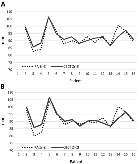
Fig. 4 Comparison of the inter-zygomatic distance measured on cone-beam computed tomography (CBCT) to values measured on PA cephalograms, stratified by operator. PA Zr-Zl: inter-zygomatic distance measured on posterior-anterior cephalograms, CBCT Zr-Zl: inter-zygomatic distance on CBCT, measured according the formula: Zr-MSP distance+Zl-MSP distance (see text). A. Operator 1. B. Operator 2.
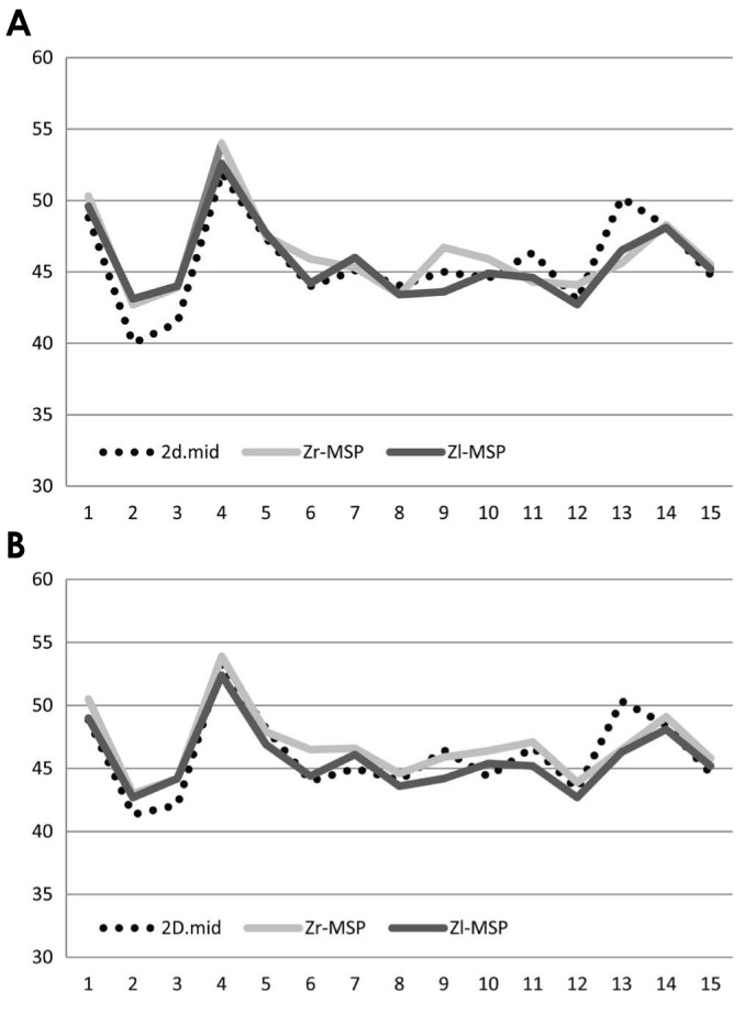
Fig. 5 Raw data of right-side and left-side cone-beam computed tomography (CBCT) measurements compared with half of the inter-zygomatic distance measured on PA cephalograms. 2D.mid=distance between the 2D mid-plane and the Zr/Zl landmarks on posterior-anterior cephalograms, given by the formula 2D Zr-Zl/2; Zr-MSP=CBCT Zr-mid-sagittal plane (MSP) distance; Zl-MSP=CBCT Zl-MSP. A. Operator 1. B. Operator 2.
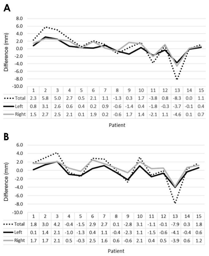
Fig. 6 Differences between measurements in millimetres, stratified by operator. A. Operator 1. B. Operator 2.
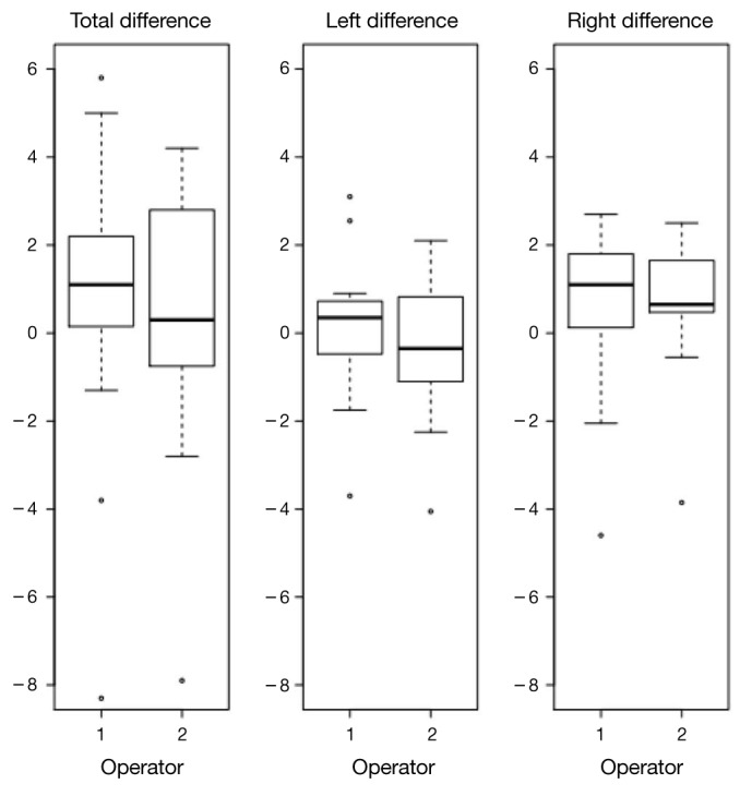
Fig. 7 Box-plots of the absolute differences show the distributions of the 3 sets of measurements assessed in this study stratified by the operator. Extreme values were measured in patient No. 13, who was considered to be an outlier and represented by dots. Most of the differences between measurements are above 0 for the total inter-zygomatic measurements and the right-side measurements, with the lower and the higher side of the box representing the first and the third quartiles of the distribution. The thick black line represents the median value, which is close to 0, indicating that the left-side measurements were more accurate than the right-side measurements.
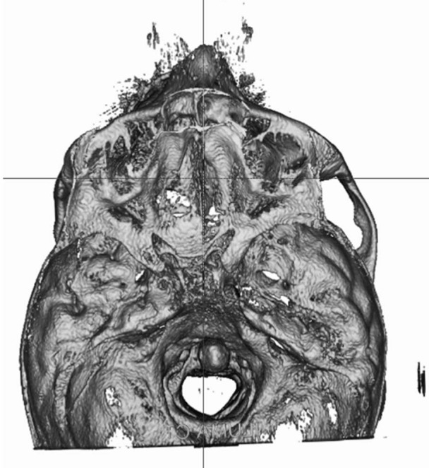
Fig. 8 Three-dimensional axial orientation of patient No. 13 showing how, given the distortion of his skull base, the mid-sagittal plane was not suitable for dividing the skull into 2 perfectly symmetric anatomical halves: measurements of this patient were considered to be outliers in the statistical analysis.
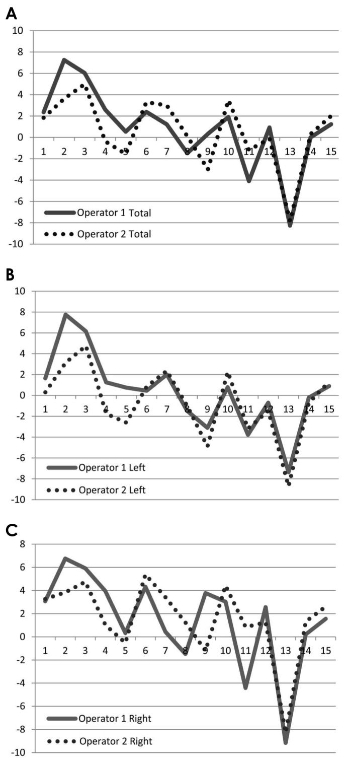
Fig. 9 Percentage difference between cone-beam computed tomography measurements and the posterior-anterior cephalogram measurements, stratified by operator (see text for the formula). A. Inter-zygomatic distance. B. Left-side measurements. C. Right-side measurements.
Reference
-
1. Mah JK, Huang JC, Choo H. Practical applications of cone-beam computed tomography in orthodontics. J Am Dent Assoc. 2010; 141(Suppl 3):7S–13S. PMID: 20884934.
Article2. Zamora N, Llamas JM, Cibrián R, Gandia JL, Paredes V. A study on the reproducibility of cephalometric landmarks when undertaking a three-dimensional (3D) cephalometric analysis. Med Oral Patol Oral Cir Bucal. 2012; 17:e678–e688. PMID: 22322503.
Article3. Langlade M. Céphalométrie orthodontique. Paris: Maloine;1978.4. Grauer D, Cevidanes LS, Proffit WR. Working with DICOM craniofacial images. Am J Orthod Dentofacial Orthop. 2009; 136:460–470. PMID: 19732681.
Article5. Olszewski R, Tanesy O, Cosnard G, Zech F, Reychler H. Reproducibility of osseous landmarks used for computed tomography based three-dimensional cephalometric analyses. J Craniomaxillofac Surg. 2010; 38:214–221. PMID: 19574058.
Article6. Nur RB, Çakan DG, Arun T. Evaluation of facial hard and soft tissue asymmetry using cone-beam computed tomography. Am J Orthod Dentofacial Orthop. 2016; 149:225–237. PMID: 26827979.
Article7. Rossini G, Cavallini C, Cassetta M, Barbato E. 3D cephalometric analysis obtained from computed tomography. Review of the literature. Ann Stomatol (Roma). 2011; 2:31–31. PMID: 22545187.8. Olszewski R, Zech F, Cosnard G, Nicolas V, Macq B, Reychler H. Three-dimensional computed tomography cephalometric craniofacial analysis: experimental validation in vitro. Int J Oral Maxillofac Surg. 2007; 36:828–833. PMID: 17825530.
Article9. Moreira CR, Sales MA, Lopes PM, Cavalcanti MG. Assessment of linear and angular measurements on three-dimensional cone-beam computed tomographic images. Oral Surg Oral Med Oral Pathol Oral Radiol Endod. 2009; 108:430–436. PMID: 19386521.
Article10. van Vlijmen OJ, Maal T, Bergé SJ, Bronkhorst EM, Katsaros C, Kuijpers-Jagtman AM. A comparison between 2D and 3D cephalometry on CBCT scans of human skulls. Int J Oral Maxillofac Surg. 2010; 39:156–160. PMID: 20044238.
Article11. Cascone P, De Ponte F, Schaerf M. Cephalometric analysis in maxillofacial surgery. A proposed computerized analysis in antero-postero projection for study of maxillofacial abnormalities. Mondo Ortod. 1987; 12:97–106. PMID: 3331722.12. Damstra J, Fourie Z, De Wit M, Ren Y. A three-dimensional comparison of a morphometric and conventional cephalometric midsagittal planes for craniofacial asymmetry. Clin Oral Investig. 2012; 16:285–294.
Article13. Hassan B, van der Stelt P, Sanderink G. Accuracy of three-dimensional measurements obtained from cone beam computed tomography surface-rendered images for cephalometric analysis: influence of patient scanning position. Eur J Orthod. 2009; 31:129–134. PMID: 19106265.
Article14. Berco M, Rigali PH Jr, Miner RM, DeLuca S, Anderson NK, Will LA. Accuracy and reliability of linear cephalometric measurements from cone-beam computed tomography scans of a dry human skull. Am J Orthod Dentofacial Orthop. 2009; 136:17.e1–17.e9. PMID: 19577142.
Article15. Periago DR, Scarfe WC, Moshiri M, Scheetz JP, Silveira AM, Farman AG. Linear accuracy and reliability of cone beam CT derived 3-dimensional images constructed using an orthodontic volumetric rendering program. Angle Orthod. 2008; 78:387–395. PMID: 18416632.
Article16. Moro A, Correra P, Boniello R, Gasparini G, Pelo S. Three-dimensional analysis in facial asymmetry: comparison with model analysis and conventional two-dimensional analysis. J Craniofac Surg. 2009; 20:417–422. PMID: 19258903.
Article17. Enlow DH, Hans MG. Essentials of facial growth. Philadelphia: WS Saunders Co.;1996.18. De Coster L. A new line of reference for the study of lateral facial teleradiographs. Am J Orthod. 1953; 39:304–306.19. Farkas LG. Anthropometry of the head and face. 2nd ed. New York: Raven Press;1994.20. Kwon TG, Park HS, Ryoo HM, Lee SH. A comparison of craniofacial morphology in patients with and without facial asymmetry - a three-dimensional analysis with computed tomography. Int J Oral Maxillofac Surg. 2006; 35:43–48. PMID: 15925488.21. Schlicher W, Nielsen I, Huang JC, Maki K, Hatcher DC, Miller AJ. Consistency and precision of landmark identification in three-dimensional cone beam computed tomography scans. Eur J Orthod. 2012; 34:263–275. PMID: 21385857.
Article22. Lagravère MO, Low C, Flores-Mir C, Chung R, Carey JP, Heo G, et al. Intraexaminer and interexaminer reliabilities of landmark identification on digitized lateral cephalograms and formatted 3-dimensional cone-beam computerized tomography images. Am J Orthod Dentofacial Orthop. 2010; 137:598–604. PMID: 20451778.
Article23. de Oliveira AE, Cevidanes LH, Phillips C, Motta A, Burke B, Tyndall D. Observer reliability of three-dimensional cephalometric landmark identification on cone-beam computerized tomography. Oral Surg Oral Med Oral Pathol Oral Radiol Endod. 2009; 107:256–265. PMID: 18718796.
Article24. Eliades T. Research methods in orthodontics: a guide to understanding orthodontic research. Heidelberg: Springer;2013.25. Gribel BF, Gribel MN, Frazão DC, McNamara JA Jr, Manzi FR. Accuracy and reliability of craniometric measurements on lateral cephalometry and 3D measurements on CBCT scans. Angle Orthod. 2011; 81:26–35. PMID: 20936951.
Article26. Neiva MB, Soares ÁC, Lisboa Cde O, Vilella Ode V, Motta AT. Evaluation of cephalometric landmark identification on CBCT multiplanar and 3D reconstructions. Angle Orthod. 2015; 85:11–17. PMID: 24713068.27. Fernandes TM, Adamczyk J, Poleti ML, Henriques JF, Friedland B, Garib DG. Comparison between 3D volumetric rendering and multiplanar slices on the reliability of linear measurements on CBCT images: an in vitro study. J Appl Oral Sci. 2015; 23:56–63. PMID: 25004053.
Article28. Cassetta M, Altieri F, Di Giorgio R, Silvestri A. Two-dimensional and three-dimensional cephalometry using cone beam computed tomography scans. J Craniofac Surg. 2015; 26:e311–e315. PMID: 26080244.29. Trpkova B, Major P, Prasad N, Nebbe B. Cephalometric landmarks identification and reproducibility: a meta analysis. Am J Orthod Dentofacial Orthop. 1997; 112:165–170. PMID: 9267228.
Article
- Full Text Links
-
- Actions
-
Cited
- CITED
-
- Close
- Share
-
- Similar articles
-
- Comparison of midsagittal reference plane in PA cephalogram and 3D CT
- A comparative study between data obtained from conventional lateral cephalometry and reconstructed three-dimensional computed tomography images
- Three-dimensional Effect of the Single Plane Proximal Femur Osteotomy
- Three-dimensional analysis of soft and hard tissue changes after mandibular setback surgery in skeletal Class III patients
- Characteristics of the pulsating jet flow through a dynamic glottal model with a lens-like constriction
