Marginal microleakage of cervical composite resin restorations bonded using etch-and-rinse and self-etch adhesives: two dimensional vs. three dimensional methods
- Affiliations
-
- 1Dental Materials Research Center and Department of Operative Dentistry, School of Dentistry, Isfahan University of Medical Sciences, Isfahan, Iran.
- 2Dental Students Research Committee and Department of Orthodontics, School of Dentistry, Isfahan University of Medical Sciences, Isfahan, Iran. ailin2214@yahoo.com
- KMID: 2163289
- DOI: http://doi.org/10.5395/rde.2016.41.2.83
Abstract
OBJECTIVES
This study was evaluated the marginal microleakage of two different adhesive systems before and after aging with two different dye penetration techniques.
MATERIALS AND METHODS
Class V cavities were prepared on the buccal and lingual surfaces of 48 human molars. Clearfil SE Bond and Single Bond (self-etching and etch-and-rinse systems, respectively) were applied, each to half of the prepared cavities, which were restored with composite resin. Half of the specimens in each group underwent 10,000 cycles of thermocycling. Microleakage was evaluated using two dimensional (2D) and three dimensional (3D) dye penetration techniques separately for each half of each specimen. Data were analyzed with SPSS 11.5 (SPSS Inc.), using the Kruskal-Wallis and Mann-Whitney U tests (α = 0.05).
RESULTS
The difference between the 2D and 3D microleakage evaluation techniques was significant at the occlusal margins of Single bond groups (p = 0.002). The differences between 2D and 3D microleakage evaluation techniques were significant at both the occlusal and cervical margins of Clearfil SE Bond groups (p = 0.017 and p = 0.002, respectively). The difference between the 2D and 3D techniques was significant at the occlusal margins of non-aged groups (p = 0.003). The difference between these two techniques was significant at the occlusal margins of the aged groups (p = 0.001). The Mann-Whitney test showed significant differences between the two techniques only at the occlusal margins in all specimens.
CONCLUSIONS
Under the limitations of the present study, it can be concluded that the 3D technique has the capacity to detect occlusal microleakage more precisely than the 2D technique.
Figure
-
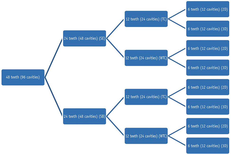
Figure 1 Classifications of specimens in the study groups depending on the type of adhesive, thermocycling and method used to assess microleakage. SB, Single Bond; SE, Clearfil SE Bond; TC, thermocycled; WTC, without thermocycling; 2D, two dimensional method; 3D, three dimensional method.
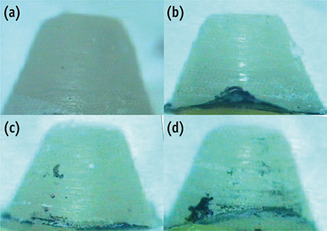
Figure 2 Examples of three dimensional specimens. (a) Score 0, no leakage; (b) Score 1, leakage depth up to one third of the internal surface; (c) Score 2, leakage depth up to two thirds of the internal surface; (d) Score 3, leakage through the entire lateral surface to the bottom of the filling.
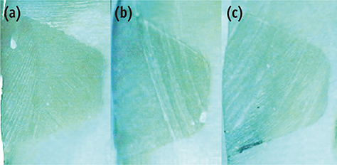
Figure 3 Examples of two dimensional specimens. (a) Score 0, no leakage; (b) Score 1, leakage depth up to one third of the internal surface; (c) Score 2, leakage depth up to two thirds of the internal surface.
Reference
-
1. Manhart J, Neuerer P, Scheibenbogen-Fuchsbrunner A, Hickel R. Three-year clinical evaluation of direct and indirect composite restorations in posterior teeth. J Prosthet Dent. 2000; 84:289–296.
Article2. Demarco FF, Corrêa MB, Cenci MS, Moraes RR, Opdam NJ. Longevity of posterior composite restorations: not only a matter of materials. Dent Mater. 2012; 28:87–101.
Article3. Opdam NJ, Loomans BA, Roeters FJ, Bronkhorst EM. Five-year clinical performance of posterior resin composite restorations placed by dental students. J Dent. 2004; 32:379–383.
Article4. Korkmaz Y, Ozel E, Attar N, Bicer CO, Firatli E. Microleakage and scanning electron microscopy evaluation of all-in-one self-etch adhesives and their respective nanocomposites prepared by erbium:yttrium-aluminum-garnet laser and bur. Lasers Med Sci. 2010; 25:493–502.
Article5. Krmek SJ, Bogdan I, Simeon P, Mehicić GP, Katanec D, Anić I. A three-dimensional evaluation of microleakage of class V cavities prepared by the very short pulse mode of the erbium:yttrium-aluminium-garnet laser. Lasers Med Sci. 2010; 25:823–828.
Article6. Waldman GL, Vaidyanathan TK, Vaidyanathan J. Microleakage and resin-to-dentin interface morphology of pre-etching versus self-etching adhesive systems. Open Dent J. 2008; 2:120–125.
Article7. Zhao XY, Li SB, Gu LJ, Li Y. Detection of marginal leakage of Class V restorations in vitro by micro-computed tomography. Oper Dent. 2014; 39:174–180.
Article8. Frankenberger R, Tay FR. Self-etch vs etch-and-rinse adhesives: effect of thermo-mechanical fatigue loading on marginal quality of bonded resin composite restorations. Dent Mater. 2005; 21:397–412.
Article9. Duarte S Jr, Dinelli W, da Silva MH. Influence of resin composite insertion technique in preparations with a high C-factor. Quintessence Int. 2007; 38:829–835.10. Geerts S, Bolette A, Seidel L, Guéders A. An in vitro evaluation of leakage of two etch and rinse and two self-etch adhesives after thermocycling. Int J Dent. 2012; 2012:852841.11. Deliperi S, Bardwell DN, Wegley C. Restoration interface microleakage using one total-etch and three self-etch adhesives. Oper Dent. 2007; 32:179–184.
Article12. Eick JD, Gwinnett AJ, Pashley DH, Robinson SJ. Current concepts on adhesion to dentin. Crit Rev Oral Biol Med. 1997; 8:306–335.
Article13. Nagpal R, Manuja N, Tyagi SP, Singh UP. In vitro bonding effectiveness of self-etch adhesives with different application techniques: a microleakage and scanning electron microscopic study. J Conserv Dent. 2011; 14:258–263.
Article14. van Dijken JW. A prospective 8-year evaluation of a mild two-step self-etching adhesive and a heavily filled two-step etch-and-rinse system in non-carious cervical lesions. Dent Mater. 2010; 26:940–946.
Article15. Van Meerbeek B, Yoshihara K, Yoshida Y, Mine A, De Munck J, Van Landuyt KL. State of the art of self-etch adhesives. Dent Mater. 2011; 27:17–28.
Article16. Proença JP, Polido M, Osorio E, Erhardt MC, Aguilera FS, García-Godoy F, Osorio R, Toledano M. Dentin regional bond strength of self-etch and total-etch adhesive systems. Dent Mater. 2007; 23:1542–1548.
Article17. Sano H, Yoshikawa T, Pereira PN, Kanemura N, Morigami M, Tagami J, Pashley DH. Long-term durability of dentin bonds made with a self-etching primer, in vivo. J Dent Res. 1999; 78:906–911.
Article18. Hayakawa T, Kikutake K, Nemoto K. Influence of self-etching primer treatment on the adhesion of resin composite to polished dentin and enamel. Dent Mater. 1998; 14:99–105.
Article19. Silveira de Araújo C, Incerti da Silva T, Ogliari FA, Meireles SS, Piva E, Demarco FF. Microleakage of seven adhesive systems in enamel and dentin. J Contemp Dent Pract. 2006; 7:26–33.
Article20. Guéders AM, Charpentier JF, Albert AI, Geerts SO. Microleakage after thermocycling of 4 etch and rinse and 3 self-etch adhesives with and without a flowable composite lining. Oper Dent. 2006; 31:450–455.
Article21. Owens BM, Johnson WW, Harris EF. Marginal permeability of self-etch and total-etch adhesive systems. Oper Dent. 2006; 31:60–67.
Article22. Stalin A, Varma BR, Jayanthi . Comparative evaluation of tensile-bond strength, fracture mode and microleakage of fifth, and sixth generation adhesive systems in primary dentition. J Indian Soc Pedod Prev Dent. 2005; 23:83–88.
Article23. Khoroushi M, Mansoori M. Marginal sealing durability of two contemporary self-etch adhesives. ISRN Dent. 2012; 2012:204813.
Article24. Hilton TJ. Can modern restorative procedures and materials reliably seal cavities? In vitro investigations. Part 2. Am J Dent. 2002; 15:279–289.25. Gwinnett JA, Tay FR, Pang KM, Wei SH. Comparison of three methods of critical evaluation of microleakage along restorative interfaces. J Prosthet Dent. 1995; 74:575–585.
Article26. Khoroushi M, Karvandi TM, Kamali B, Mazaheri H. Marginal microleakage of resin-modified glass-ionomer and composite resin restorations: effect of using etch-and-rinse and self-etch adhesives. Indian J Dent Res. 2012; 23:378–383.
Article27. Pashley DH, Tay FR. Aggressiveness of contemporary self-etching adhesives. Part II: etching effects on unground enamel. Dent Mater. 2001; 17:430–444.
Article28. De Munck J, Vargas M, Iracki J, Van Landuyt K, Poitevin A, Lambrechts P, Van Meerbeek B. One-day bonding effectiveness of new self-etch adhesives to bur-cut enamel and dentin. Oper Dent. 2005; 30:39–49.29. De Munck J, Van Landuyt K, Peumans M, Poitevin A, Lambrechts P, Braem M, Van Meerbeek B. A critical review of the durability of adhesion to tooth tissue: methods and results. J Dent Res. 2005; 84:118–132.
Article30. Doerr CL, Hilton TJ, Hermesch CB. Effect of thermocycling on the microleakage of conventional and resin-modified glass ionomers. Am J Dent. 1996; 9:19–21.31. Yap AU. Effects of storage, thermal and load cycling on a new reinforced glass-ionomer cement. J Oral Rehabil. 1998; 25:40–44.
Article32. Koshiro K, Inoue S, Tanaka T, Koase K, Fujita M, Hashimoto M, Sano H. In vivo degradation of resin-dentin bonds produced by a self-etch vs. a total-etch adhesive system. Eur J Oral Sci. 2004; 112:368–375.
Article33. Koshiro K, Inoue S, Sano H, De Munck J, Van Meerbeek B. In vivo degradation of resin-dentin bonds produced by a self-etch and an etch-and-rinse adhesive. Eur J Oral Sci. 2005; 113:341–348.
Article
- Full Text Links
-
- Actions
-
Cited
- CITED
-
- Close
- Share
-
- Similar articles
-
- Microleakage and characteristics of resin-tooth tissues interface of a selfetch and an etch-and-rinse adhesive systems
- Effect of various bleaching treatments on shear bond strength of different universal adhesives and application modes
- Micro-CT evaluation of internal adaptation in resin fillings with different dentin adhesives
- The effect of various bonding systems on the microtensile bond strength of immediate and delayed dentin sealing
- Enamel adhesion of light- and chemical-cured composites coupled by two step self-etch adhesives
