Yonsei Med J.
2013 Jul;54(4):927-934. 10.3349/ymj.2013.54.4.927.
Clinical and Radiological Characteristics of 2009 H1N1 Influenza Associated Pneumonia in Young Male Adults
- Affiliations
-
- 1Department of Internal Medicine, Armed Forces Capital Hospital, Seongnam, Korea.
- 2Department of Pulmonary and Critical Care Medicine, and Clinical Research Center for Chronic Obstructive Airway Diseases, Asan Medical Center, University of Ulsan College of Medicine, Seoul, Korea. seiwon@amc.seoul.kr
- KMID: 2158228
- DOI: http://doi.org/10.3349/ymj.2013.54.4.927
Abstract
- PURPOSE
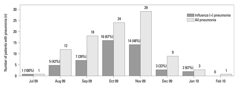
Pneumonia was an important cause of death in 2009 H1N1 influenza pandemic (pH1N1). Clinical characteristics of pH1N1 have been described well, but discriminative characteristics suggesting pH1N1 infection in pneumonia patients are not evident today. We evaluated differences between clinical and radiologic characteristics for those associated and not associated with pH1N1 influenza during the pandemic period.
MATERIALS AND METHODS
We reviewed all patients with pneumonia who visited the Armed Forces Capital Hospital between July 2009 and February 2010. During this period, all pneumonia patients were tested for pH1N1 by reverse transcription-polymerase chain reaction (RT-PCR) using nasopharyngeal specimens.
RESULTS
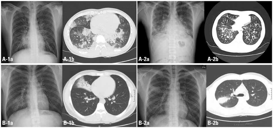
In total, 98 patients with pneumonia were enrolled. Their median age was 20 years and all patients were males. Forty-nine (50%) of patients had pH1N1 infection and the others (50%) had negative results in pH1N1 RT-PCR. Patients with pH1N1 infection complained of dyspnea more commonly (83.3% vs. 29.0%; p<0.001), had higher Acute Physiology and Chronic Health Evaluation (APACHE) II scores [5 (range, 0-12) vs. 3 (range, 0-11); p<0.01], fewer days of prehospital illness [2 (range, 0-10) vs. 4 (range, 0-14); p=0.001], and a higher chance of bilateral infiltrates on chest X-ray (CXR) (67.3% vs. 14.3%; p<0.001) and ground-glass opacity (GGO) lesions on computed tomography (CT; 48.9% vs. 22.0%; p<0.001) than patients without pH1N1 infection.
CONCLUSION
Dyspnea, bilateral infiltrates on CXR, and GGO on CT were dominant features in pH1N1-associated pneumonia. Understanding these characteristics can help selection of patients who require prompt antiviral therapy.
MeSH Terms
-
Adolescent
Adult
Antiviral Agents/therapeutic use
Dyspnea/virology
Humans
Influenza A Virus, H1N1 Subtype/genetics/*pathogenicity
Influenza, Human/*complications/radiography/virology
Male
Middle Aged
Pneumonia/etiology/radiography
Pneumonia, Viral/drug therapy/etiology/*radiography/*virology
Radiography, Thoracic
Tomography, X-Ray Computed
Young Adult
Antiviral Agents
Figure
Reference
-
1. Centers for Disease Control and Prevention (CDC). Update: infections with a swine-origin influenza A (H1N1) virus--United States and other countries, April 28, 2009. MMWR Morb Mortal Wkly Rep. 2009; 58:431–433.2. Centers for Disease Control and Prevention (CDC). Swine influenza A (H1N1) infection in two children--Southern California, March-April 2009. MMWR Morb Mortal Wkly Rep. 2009; 58:400–402.3. Centers for Disease Control and Prevention (CDC). Update: swine influenza A (H1N1) infections--California and Texas, April 2009. MMWR Morb Mortal Wkly Rep. 2009; 58:435–437.4. Kim WJ. Pandemic influenza (H1N1 2009): experience and lessons. Infect Chemother. 2010; 42:61–63.
Article5. Jain S, Kamimoto L, Bramley AM, Schmitz AM, Benoit SR, Louie J, et al. Hospitalized patients with 2009 H1N1 influenza in the United States, April-June 2009. N Engl J Med. 2009; 361:1935–1944.
Article6. Viasus D, Paño-Pardo JR, Pachón J, Riera M, López-Medrano F, Payeras A, et al. Timing of oseltamivir administration and outcomes in hospitalized adults with pandemic 2009 influenza A(H1N1) virus infection. Chest. 2011; 140:1025–1032.
Article7. Cunha BA. Swine Influenza (H1N1) pneumonia: clinical considerations. Infect Dis Clin North Am. 2010; 24:203–228.
Article8. Riquelme R, Riquelme M, Rioseco ML, Inzunza C, Gomez Y, Contreras C, et al. Characteristics of hospitalised patients with 2009 H1N1 influenza in Chile. Eur Respir J. 2010; 36:864–869.
Article9. Agarwal PP, Cinti S, Kazerooni EA. Chest radiographic and CT findings in novel swine-origin influenza A (H1N1) virus (S-OIV) infection. AJR Am J Roentgenol. 2009; 193:1488–1493.
Article10. Cunha BA, Pherez FM, Strollo S. Swine influenza (H1N1): diagnostic dilemmas early in the pandemic. Scand J Infect Dis. 2009; 41:900–902.
Article11. Rello J, Rodríguez A, Ibañez P, Socias L, Cebrian J, Marques A, et al. Intensive care adult patients with severe respiratory failure caused by Influenza A (H1N1)v in Spain. Crit Care. 2009; 13:R148.
Article12. Denholm JT, Gordon CL, Johnson PD, Hewagama SS, Stuart RL, Aboltins C, et al. Hospitalised adult patients with pandemic (H1N1) 2009 influenza in Melbourne, Australia. Med J Aust. 2010; 192:84–86.13. Louie JK, Acosta M, Winter K, Jean C, Gavali S, Schechter R, et al. Factors associated with death or hospitalization due to pandemic 2009 influenza A(H1N1) infection in California. JAMA. 2009; 302:1896–1902.
Article14. Kumar A, Zarychanski R, Pinto R, Cook DJ, Marshall J, Lacroix J, et al. Critically ill patients with 2009 influenza A(H1N1) infection in Canada. JAMA. 2009; 302:1872–1879.
Article15. Soto-Abraham MV, Soriano-Rosas J, Díaz-Quiñónez A, Silva-Pereyra J, Vazquez-Hernandez P, Torres-López O, et al. Pathological changes associated with the 2009 H1N1 virus. N Engl J Med. 2009; 361:2001–2003.
Article16. Novel Swine-Origin Influenza A (H1N1) Virus Investigation Team. Dawood FS, Jain S, Finelli L, Shaw MW, Lindstrom S, et al. Emergence of a novel swine-origin influenza A (H1N1) virus in humans. N Engl J Med. 2009; 360:2605–2615.
Article17. Chowell G, Bertozzi SM, Colchero MA, Lopez-Gatell H, Alpuche-Aranda C, Hernandez M, et al. Severe respiratory disease concurrent with the circulation of H1N1 influenza. N Engl J Med. 2009; 361:674–679.
Article18. Riquelme R, Torres A, Rioseco ML, Ewig S, Cillóniz C, Riquelme M, et al. Influenza pneumonia: a comparison between seasonal influenza virus and the H1N1 pandemic. Eur Respir J. 2011; 38:106–111.
Article19. World Health Organization. WHO guidelines for pharmacological management of pandemic (H1N1) 2009 influenza and other influenza viruses. 2010. accessed on 2011 July 15. Geneva: Available at: http://www.who.int/csr/resources/publications/swineflu/h1n1_use_antivirals_20090820/en/index.html.20. Mori T, Morii M, Terada K, Wada Y, Kuroiwa Y, Hotsubo T, et al. Clinical characteristics and computed tomography findings in children with 2009 pandemic influenza A (H1N1) viral pneumonia. Scand J Infect Dis. 2011; 43:47–54.
Article21. Ajlan AM, Quiney B, Nicolaou S, Müller NL. Swine-origin influenza A (H1N1) viral infection: radiographic and CT findings. AJR Am J Roentgenol. 2009; 193:1494–1499.
Article22. Mollura DJ, Asnis DS, Crupi RS, Conetta R, Feigin DS, Bray M, et al. Imaging findings in a fatal case of pandemic swine-origin influenza A (H1N1). AJR Am J Roentgenol. 2009; 193:1500–1503.
Article23. Remy-Jardin M, Remy J, Giraud F, Wattinne L, Gosselin B. Computed tomography assessment of ground-glass opacity: semiology and significance. J Thorac Imaging. 1993; 8:249–264.24. Remy-Jardin M, Giraud F, Remy J, Copin MC, Gosselin B, Duhamel A. Importance of ground-glass attenuation in chronic diffuse infiltrative lung disease: pathologic-CT correlation. Radiology. 1993; 189:693–698.
Article25. John SD, Ramanathan J, Swischuk LE. Spectrum of clinical and radiographic findings in pediatric mycoplasma pneumonia. Radiographics. 2001; 21:121–131.
Article26. King MA, Pope-Harman AL, Allen JN, Christoforidis GA, Christoforidis AJ. Acute eosinophilic pneumonia: radiologic and clinical features. Radiology. 1997; 203:715–719.
Article27. Perez-Padilla R, de la Rosa-Zamboni D, Ponce de Leon S, Hernandez M, Quiñones-Falconi F, Bautista E, et al. Pneumonia and respiratory failure from swine-origin influenza A (H1N1) in Mexico. N Engl J Med. 2009; 361:680–689.
Article28. Ohrui T, Takahashi H, Ebihara S, Matsui T, Nakayama K, Sasaki H. Influenza A virus infection and pulmonary microthromboembolism. Tohoku J Exp Med. 2000; 192:81–86.
Article29. Choi WI, Yim JJ, Park J, Kim SC, Na MJ, Lee WY, et al. Clinical characteristics and outcomes of H1N1-associated pneumonia among adults in South Korea. Int J Tuberc Lung Dis. 2011; 15:270–275. i30. Louie JK, Acosta M, Jamieson DJ, Honein MA. California Pandemic (H1N1) Working Group. Severe 2009 H1N1 influenza in pregnant and postpartum women in California. N Engl J Med. 2010; 362:27–35.
Article31. Jamieson DJ, Honein MA, Rasmussen SA, Williams JL, Swerdlow DL, Biggerstaff MS, et al. H1N1 2009 influenza virus infection during pregnancy in the USA. Lancet. 2009; 374:451–458.
Article32. Jung JY, Park MS, Kim YS, Park BH, Kim SK, Chang J, et al. Healthcare-associated pneumonia among hospitalized patients in a Korean tertiary hospital. BMC Infect Dis. 2011; 11:61.
Article
- Full Text Links
- Actions
-
Cited
- CITED
-
- Close
- Share
- Similar articles
-
- Influenza Associated Pneumonia
- Epidemiology, clinical manifestations, and management of pandemic novel Influenza A (H1N1)
- A Case of Pseudomembranous Tracheobronchitis Complicated by Coinfection of 2009 Pandemic Influenza A/H1N1 and Staphylococcus aureus
- 2009 Pandemic Influenza A(H1N1) Infections in the Pediatric Cancer Patients and Comparative Analysis with Seasonal Influenza
- Novel Influenza A/H1N1 Pandemic: Current Status and Prospects


