Yonsei Med J.
2008 Oct;49(5):843-850. 10.3349/ymj.2008.49.5.843.
In Utero Exposure to 2,3,7,8-Tetrachlorodibenzo-p-Dioxin Affects the Development of Reproductive System in Mouse
- Affiliations
-
- 1Department of Urology and the Urological Science Institute, Yonsei University College of Medicine, Seoul, Korea. swhan@yuhs.ac
- 2Brain Korea 21 Project for Medical Science, Yonsei University College of Medicine, Seoul, Korea.
- KMID: 2158204
- DOI: http://doi.org/10.3349/ymj.2008.49.5.843
Abstract
- PURPOSE
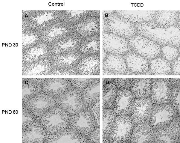
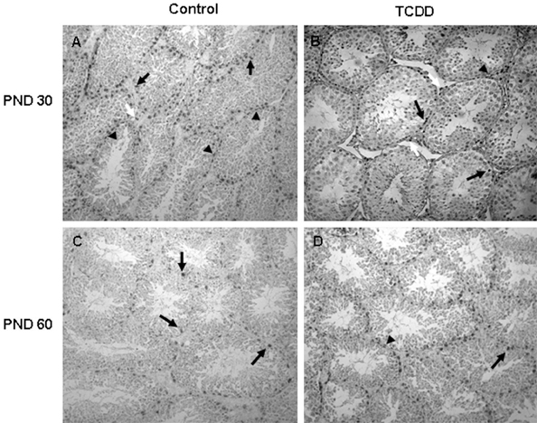
Exposure of male reproductive organs to 2,3,7,8-Tetrachlorodibenzo-p-Dioxin (TCDD) has been reported to cause developmental changes. In this study, we evaluated the effects of in utero TCDD exposure on male reproductive development. MATERIALS AND METHODS: Pregnant C57BL/6 mice were administered a single intraperitoneal injection of TCDD (1microgram/kg) on gestation day (GD) 15. The offspring were examined in the immature stage on postnatal day (PND) 30 and in the mature stage on PND 60. The testes were examined for histological changes, androgen receptor (AR), proliferating cell nuclear antigen (PCNA) and apoptosis following the measurement of morphological changes. RESULTS: Anogenital distance (AGD) and testis weights were reduced by TCDD exposure both on PND 30 and PND 60 while body weights and length of male offspring were not affected by TCDD. The regular sperm developmental stage was impaired with TCDD treatment on PND 30. However, no difference was found between the control group and TCDD groups on PND 60. Simultaneously, the expression of AR was also reduced on PND 30, while it was increased on PND 60 compared with the control group. The expression of PCNA was decreased whereas apoptosis was not affected by TCDD both on PND 30 and PND 60. CONCLUSION: These results suggest that in utero exposure to TCDD influences the development of testes by inhibiting the expression of AR and PCNA. Moreover, the adverse effects of TCDD on male offspring reduced over time.
MeSH Terms
-
Animals
Apoptosis/drug effects
Cell Proliferation
Embryonic Development/*drug effects
Environmental Pollutants/*toxicity
Female
Male
*Maternal Exposure
Mice
Mice, Inbred C57BL
Organ Size/drug effects
Pregnancy
Receptors, Androgen/metabolism
Testis/*drug effects/embryology/pathology
Tetrachlorodibenzodioxin/*toxicity
Figure
Reference
-
1. Chia SE. Endocrine disruptors and male reproductive function--a short review. Int J Androl. 2000. 3:45–46.2. Schecter A, Cramer P, Boggess K, Stanley J, Päpke O, Olson J, et al. Intake of dioxins and related compounds from food in the U.S. population. J Toxicol Environ Health A. 2001. 63:1–18.
Article3. Safe SH. Comparative toxicology and mechanism of action of polychlorinated dibenzo-p-dioxins and dibenzofurans. Annu Rev Pharmacol Toxicol. 1986. 26:371–399.
Article4. Fingerhut MA, Halperin WE, Marlow DA, Piacitelli LA, Honchar PA, Sweeney MH, et al. Cancer mortality in workers exposed to 2,3,7,8-tetrachlorodibenzo-p-dioxin. N Engl J Med. 1991. 324:212–218.
Article5. Goodman DG, Sauer RM. Hepatotoxicity and carcinogenicity in female Sprague-Dawley rats treated with 2,3,7,8-tetrachlorodibenzo-p-dioxin (TCDD): a pathology working group reevaluation. Regul Toxicol Pharmacol. 1992. 15:245–252.
Article6. Birnbaum LS, Harris MW, Stocking LM, Clark AM, Morrissey RE. Retinoic acid and 2,3,7,8-tetrachlorodibenzo-p-dioxin selectively enhance teratogenesis in C57BL/6N mice. Toxicol Appl Pharmacol. 1989. 98:487–500.
Article7. Faqi AS, Dalsenter PR, Merker HJ, Chahoud I. Reproductive toxicity and tissue concentrations of low doses of 2,3,7,8-tetrachlorodibenzo-p-dioxin in male offspring rats exposed throughout pregnancy and lactation. Toxicol Appl Pharmacol. 1998. 150:383–392.
Article8. Mably TA, Moore RW, Goy RW, Peterson RE. In utero and lactational exposure of male rats to 2,3,7,8-tetrachlorodibenzo-p-dioxin. 2. Effects on sexual behavior and the regulation of luteinizing hormone secretion in adulthood. Toxicol Appl Pharmacol. 1992. 114:108–117.
Article9. Mably TA, Bjerke DL, Moore RW, Gendron-Fitzpatrick A, Peterson RE. In utero and lactational exposure of male rats to 2,3,7,8-tetrachlorodibenzo-p-dioxin. 3. Effects on spermatogenesis and reproductive capability. Toxicol Appl Pharmacol. 1992. 114:118–126.
Article10. Theobald HM, Peterson RE. In utero and lactational exposure to 2,3,7,8-tetrachlorodibenzo-rho-dioxin: effects on development of the male and female reproductive system of the mouse. Toxicol Appl Pharmacol. 1997. 145:124–135.
Article11. Mooradian AD, Morley JE, Korenman SG. Biological actions of androgens. Endocr Rev. 1987. 8:1–28.
Article12. Bjerke DL, Peterson RE. Reproductive toxicity of 2,3,7,8-tetrachlorodibenzo-p-dioxin in male rats: different effects of In utero versus lactational exposure. Toxicol Appl Pharmacol. 1994. 127:241–249.
Article13. Ohsako S, Miyabara Y, Nishimura N, Kurosawa S, Sakaue M, Ishimura R, et al. Maternal exposure to a low dose of 2,3,7,8-tetrachlorodibenzo-p-dioxin (TCDD) suppressed the development of reproductive organs of male rats: dose-dependent increase of mRNA levels of 5alpha-reductase type 2 in contrast to decrease of androgen receptor in the pubertal ventral prostate. Toxicol Sci. 2001. 60:132–143.14. Ohsako S, Miyabara Y, Sakaue M, Ishimura R, Kakeyama M, Izumi H, et al. Developmental stage-specific effects of perinatal 2,3,7,8-tetrachlorodibenzo-p-dioxin exposure on reproductive organs of male rat offspring. Toxicol Sci. 2002. 66:283–292.15. Theobald HM, Roman BL, Lin TM, Ohtani S, Chen SW, Peterson RE. 2,3,7,8-tetrachlorodibenzo-p-dioxin inhibits luminal cell differentiation and androgen responsiveness of the ventral prostate without inhibiting prostatic 5alpha-dihydrotestosterone formation or testicular androgen production in rat offspring. Toxicol Sci. 2000. 58:324–338.16. de Kretser DM, Loveland KL, Meinhardt A, Simorangkir D, Wreford N. Spermatogenesis. Hum Reprod. 1998. 13:Suppl 1. 1–8.
Article17. Rodriguez I, Ody C, Araki K, Garcia I, Vassalli P. An early and massive wave of germinal cell apoptosis is required for the development of functional spermatogenesis. EMBO J. 1997. 16:2262–2270.
Article18. Richburg JH, Boekelheide K. Mono-(2-ethylhexyl) phthalate rapidly alters both Sertoli cell vimentin filaments and germ cell apoptosis in young rat testes. Toxicol Appl Pharmacol. 1996. 137:42–50.
Article19. Nandi S, Banerjee PP, Zirkin BR. Germ cell apoptosis in the testes of Sprague Dawley rats following testosterone withdrawal by ethane 1,2-dimethanesulfonate administration: relationship to Fas? Biol Reprod. 1999. 61:70–75.
Article20. Kim IH, Son HY, Cho SW, Ha CS, Kang BH. Zearalenone induces male germ cell apoptosis in rats. Toxicol Lett. 2003. 138:185–192.
Article21. Schultz R, Suominen J, Värre T, Hakovirta H, Parvinen M, Toppari J, et al. Expression of aryl hydrocarbon receptor and aryl hydrocarbon receptor nuclear translocator messenger ribonucleic acids and proteins in rat and human testis. Endocrinology. 2003. 144:767–776.
Article22. Bravo R. Synthesis of the nuclear protein cyclin (PCNA) and its relationship with DNA replication. Exp Cell Res. 1986. 163:287–293.
Article23. Bravo R, Frank R, Blundell PA, Macdonald-Bravo H. Cyclin/PCNA is the auxiliary protein of DNA polymerase-delta. Nature. 1987. 326:515–517.24. Morris GF, Mathews MB. Regulation of proliferating cell nuclear antigen during the cell cycle. J Biol Chem. 1989. 264:13856–13864.
Article25. Schlatt S, Weinbauer GF. Immunohistochemical localization of proliferating cell nuclear antigen as a tool to study cell proliferation in rodent and primate testes. Int J Androl. 1994. 17:214–222.
Article26. Gray LE Jr, Kelce WR, Monosson E, Ostby JS, Birnbaum LS. Exposure to TCDD during development permanently alters reproductive function in male Long Evans rats and hamsters: reduced ejaculated and epididymal sperm numbers and sex accessory gland weights in offspring with normal androgenic status. Toxicol Appl Pharmacol. 1995. Mar. 131:108–118.27. el-Sabeawy F, Wang S, Overstreet J, Miller M, Lasley B, Enan E. Treatment of rats during pubertal development with 2,3,7,8-tetrachlorodibenzo-p-dioxin alters both signaling kinase activities and epidermal growth factor receptor binding in the testis and the motility and acrosomal reaction of sperm. Toxicol Appl Pharmacol. 1998. 150:427–442.
Article28. Cooke GM, Price CA, Oko RJ. Effects of In utero and lactational exposure to 2,3,7,8-tetrachlorodibenzo-p-dioxin (TCDD) on serum androgens and steroidogenic enzyme activities in the male rat reproductive tract. J Steroid Biochem Mol Biol. 1998. 67:347–354.
Article29. Chang C, Saltzman A, Yeh S, Young W, Keller E, Lee HJ, et al. Androgen receptor: an overview. Crit Rev Eukaryot Gene Expr. 1995. 5:97–125.
Article30. McIntyre BS, Barlow NJ, Foster PM. Male rats exposed to linuron In utero exhibit permanent changes in anogenital distance, nipple retention, and epididymal malformations that result in subsequent testicular atrophy. Toxicol Sci. 2002. 65:62–70.
Article31. Hotchkiss AK, Parks-Saldutti LG, Ostby JS, Lambright C, Furr F, Vandenbergh JG, et al. A mixture of the "antiandrogens" linuron and butyl benzyl phthalate alters sexual differentiation of the male rat in a cumulative fashion. Biol Reprod. 2004. 71:1852–1861.
Article32. Scott HM, Hutchison GR, Mahood IK, Hallmark N, Welsh M, Gendt KD, et al. Role of androgens in fetal testis development and dysgenesis. Endocrinology. 2007. 148:2027–2036.
Article33. Hamm JT, Sparrow BR, Wolf D, Birnbaum LS. In utero and lactational exposure to 2,3,7,8-tetrachlorodibenzo-p-dioxin alters postnatal development of seminal vesicle epithelium. Toxicol Sci. 2000. 54:424–430.
Article34. Pirkle JL, Wolfe WH, Patterson DG, Needham LL, Michalek JE, Miner JC, et al. Estimates of the half-life of 2,3,7,8-tetrachlorodibenzo-p-dioxin in Vietnam Veterans of Operation Ranch Hand. J Toxicol Environ Health. 1989. 27:165–171.
Article35. Perry JE, Tindall DJ. Androgens regulate the expression of proliferating cell nuclear antigen posttranscriptionally in the human prostate cancer cell line, LNCaP. Cancer Res. 1996. 56:1539–1544.
- Full Text Links
- Actions
-
Cited
- CITED
-
- Close
- Share
- Similar articles
-
- Teratological effect of 2,3,7,8-tetrachlorodibenzo-p-dioxin (TCDD): induction of cleft palate in the ddY and C57BL/6 mouse
- Dioxins and Health: Human Exposure Level and Epidemiologic Evidences of Health Effects
- A Study on the Correlation between Categorization of the Individual Exposure Levels to Agent Orange and Serum Dioxin Levels Among the Korean Vietnam Veterans
- Cryopreservation of mouse resources
- A Case of Incidental Epidermolytic Hyperkeratosis Occurring Normal Looking Skin Adjacent to Folliculitic Papules: In Veterans Who Participated in Vietnam War


