Ann Dermatol.
2011 Sep;23(Suppl 1):S20-S24. 10.5021/ad.2011.23.S1.S20.
CK20 Positive Large-cell Neuroendocrine Carcinoma Presenting with Skin Metastases
- Affiliations
-
- 1Department of Dermatology, School of Medicine, Kyung Hee University, Seoul, Korea. nikim@khmc.or.kr
- KMID: 2156743
- DOI: http://doi.org/10.5021/ad.2011.23.S1.S20
Abstract
- We present a case of cytokeratin (CK) 20-positive large cell neuroendocrine carcinoma (LCNEC) presenting with multiple skin metastases as the primary manifestation. The patient was a 55-year-old man who presented with a one- month history of subcutaneous skin colored nodules of various sizes on his trunk. Pathologic examination of the skin revealed a nested and solid proliferation of large undifferentiated cells with vesicular nuclei and prominent nucleoli. Tumor cells were found to be immunohistochemically positive for CK 20, chromogranin A, synaptophysin, and CD56. Based on these features, the tumor was diagnosed as a large cell neuroendocrine carcinoma with multiple skin metastases. Computed tomographic (CT) imaging found metastatic foci in the liver, pleura, bone, and lymph nodes. We were unable to identify the primary site of origin. To the best of our knowledge, this is the first case of a large cell neuroendocrine carcinoma with a primary manifestation of multiple skin metastases.
MeSH Terms
Figure
-

Fig. 1 Skin examination revealed multiple well-demarcated, skin-colored subcutaneous nodules on the trunk.

Fig. 2 Microscopic findings of a large cell neuroendocrine carcinoma. (A) Low-power view showing sheets of tumor cells with a trabecular or organoid growth pattern in the subcutaneous area. (B) High-power view showing large pleomorphic cells with hyperchromatic nuclei, coarse chromatin, and numerous mitotic figures (H&E, A: ×40, B: ×200, respectively).
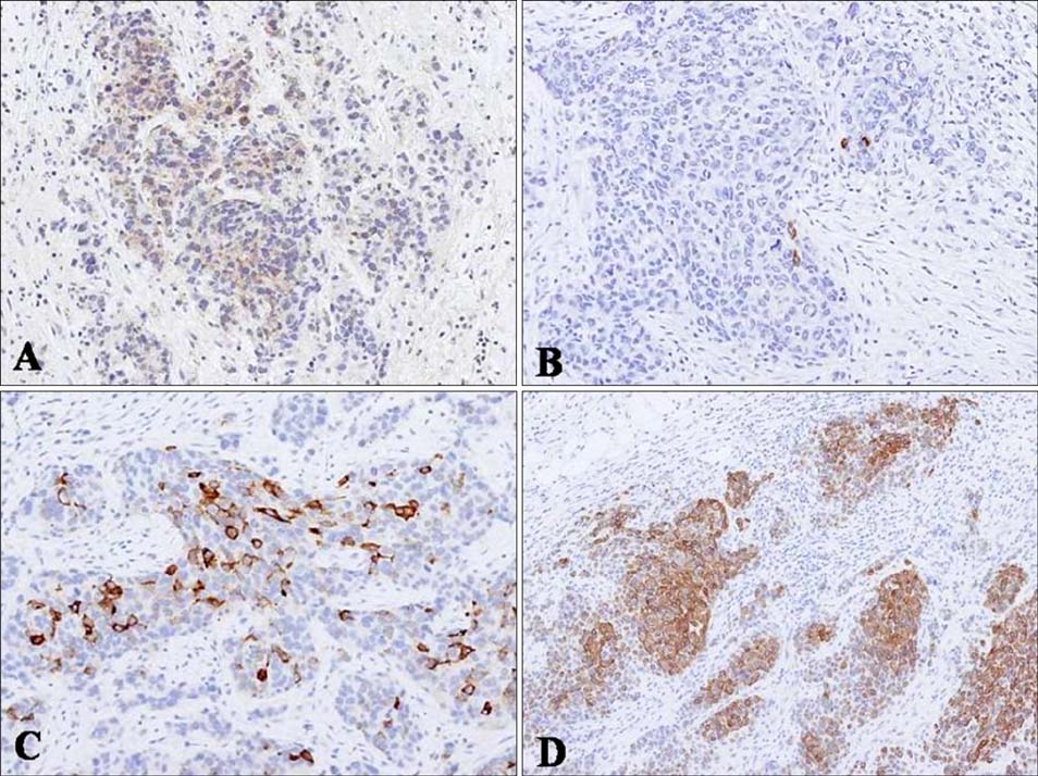
Fig. 3 (A) Immunohistochemical staining for CD56 showed positivity for tumor cells in a cytoplasmic pattern. (B, C) Tumor cells were focally positive for chromogranin and synaptophysin. (D) CK20, showing diffuse cytoplasmic staining of tumor cells (Immunoperoxidase, ×100).
Reference
-
1. Travis WD, Linnoila RI, Tsokos MG, Hitchcock CL, Cutler GB Jr, Nieman L, et al. Neuroendocrine tumors of the lung with proposed criteria for large-cell neuroendocrine carcinoma. An ultrastructural, immunohistochemical, and flow cytometric study of 35 cases. Am J Surg Pathol. 1991. 15:529–553.
Article2. Chu P, Wu E, Weiss LM. Cytokeratin 7 and cytokeratin 20 expression in epithelial neoplasms: a survey of 435 cases. Mod Pathol. 2000. 13:962–972.
Article3. Jiang SX, Kameya T, Shoji M, Dobashi Y, Shinada J, Yoshimura H. Large cell neuroendocrine carcinoma of the lung: a histologic and immunohistochemical study of 22 cases. Am J Surg Pathol. 1998. 22:526–537.4. Okoshi K, Saiga T, Hisamori S, Iwaisako K, Kobayashi H, Ogawa H. A case of cytokeratin 20-positive large-cell neuroendocrine carcinoma of the breast. Breast Cancer. 2009. [Epub ahead of print].
Article5. Wang KL, Yang YC, Wang TY, Chen JR, Chen TC, Chen HS, et al. Neuroendocrine carcinoma of the uterine cervix: a clinicopathologic retrospective study of 31 cases with prognostic implications. J Chemother. 2006. 18:209–216.
Article6. Sato K, Waseda R, Tatsuzawa Y, Fujinaga H, Wakabayashi T, Ueda Y, et al. Composite large cell neuroendocrine carcinoma and adenocarcinoma of the common bile duct. J Clin Pathol. 2006. 59:105–107.
Article7. Dundr P, Pesl M, Povýsil C, Vítková I, Dvorácek J. Large cell neuroendocrine carcinoma of the urinary bladder with lymphoepithelioma-like features. Pathol Res Pract. 2003. 199:559–563.
Article8. Veras E, Deavers MT, Silva EG, Malpica A. Ovarian nonsmall cell neuroendocrine carcinoma: a clinicopathologic and immunohistochemical study of 11 cases. Am J Surg Pathol. 2007. 31:774–782.9. Grabowski P, Schönfelder J, Ahnert-Hilger G, Foss HD, Heine B, Schindler I, et al. Expression of neuroendocrine markers: a signature of human undifferentiated carcinoma of the colon and rectum. Virchows Arch. 2002. 441:256–263.
Article10. Lee WJ, Kim CH, Chang SE, Lee MW, Choi JH, Moon KC, et al. Cutaneous metastasis from large-cell neuroendocrine carcinoma of the urinary bladder expressing CK20 and TTF-1. Am J Dermatopathol. 2009. 31:166–169.
Article11. Lee WJ, Oh SH, Chang SE, Lee MW, Choi JH, Moon KC, et al. Skin metastasis of neuroendocrine carcinoma arising in the rectum. Ann Dermatol. 2007. 19:163–165.
Article12. Miettinen M. Keratin 20: immunohistochemical marker for gastrointestinal, urothelial, and Merkel cell carcinomas. Mod Pathol. 1995. 8:384–388.13. Ordóñez NG. Thyroid transcription factor-1 is a marker of lung and thyroid carcinomas. Adv Anat Pathol. 2000. 7:123–127.
Article14. Pulitzer MP, Amin BD, Busam KJ. Merkel cell carcinoma: review. Adv Anat Pathol. 2009. 16:135–144.15. Nassar H, Albores-Saavedra J, Klimstra DS. High-grade neuroendocrine carcinoma of the ampulla of vater: a clinicopathologic and immunohistochemical analysis of 14 cases. Am J Surg Pathol. 2005. 29:588–594.16. Adegbola T, Connolly CE, Mortimer G. Small cell neuroendocrine carcinoma of the breast: a report of three cases and review of the literature. J Clin Pathol. 2005. 58:775–778.
Article17. Kato T, Terashima T, Tomida S, Yamaguchi T, Kawamura H, Kimura N, et al. Cytokeratin 20-positive large cell neuroendocrine carcinoma of the colon. Pathol Int. 2005. 55:524–529.
Article18. Dundr P, Fischerová D, Povýsil C, Cibula D. Primary pure large-cell neuroendocrine carcinoma of the ovary. Pathol Res Pract. 2008. 204:133–137.
Article19. Kasami M, Muramatsu K, Kawahata K, Yoshikawa S, Kiyohara Y. Large-cell neuroendocrine carcinoma of the skin, with lymphoid stroma. Am J Dermatopathol. 2007. 29:578–580.
Article
- Full Text Links
-
- Actions
-
Cited
- CITED
-
- Close
- Share
-
- Similar articles
-
- CK20 Negative and CK7 Positive Merkel Cell Carcinoma of the Conjunctiva: Brief Case Report
- Skin Metastasis of Neuroendocrine Carcinoma Arising in the Rectum
- A Case of Large Cell Neuroendocrine Carcinoma of the Colon
- Metastatic Large Cell Neuroendocrine Carcinoma of the Lung Mimicking a Merkel Cell Carcinoma
- Two Cases of Neuroendocrine Carcinomas of the Stomach: Large Cell Carcinoma and Small Cell Carcinoma
