Ann Dermatol.
2009 Nov;21(4):399-401. 10.5021/ad.2009.21.4.399.
A Case of Syphilitic Keratoderma Concurrent with Syphilitic Uveitis
- Affiliations
-
- 1Department of Dermatology, Busan Paik Hospital, College of Medicine, Inje University, Busan, Korea. paikderma@hanmail.net
- 2Department of Ophthalmology, Busan Paik Hospital, College of Medicine, Inje University, Busan, Korea.
- KMID: 2156505
- DOI: http://doi.org/10.5021/ad.2009.21.4.399
Abstract
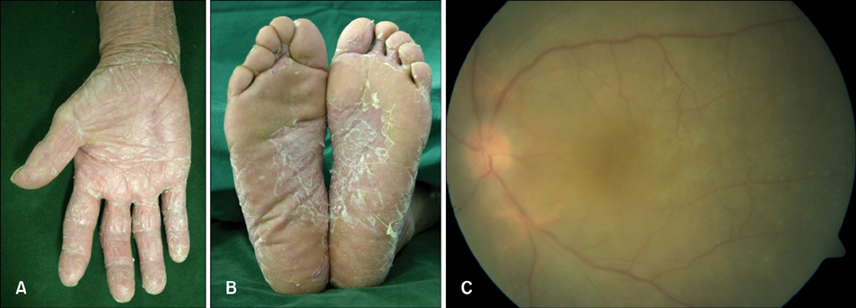
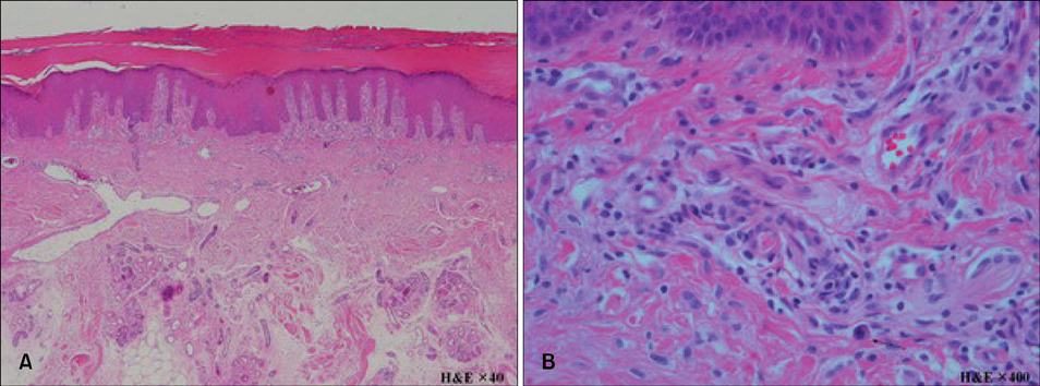
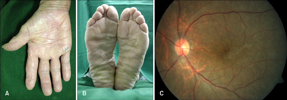
- Syphilitic keratoderma is a rare cutaneous manifestation of secondary syphilis, characterized by symmetrical and diffuse hyperkeratosis of the palms and soles. In addition, no cases of syphilitic keratoderma and uveitis have been reported in the dermatologic literature. A 69-year-old woman presented with steroid-resistant hyperkeratotic patches on the palms and soles and uveitis for 4 months. As steroid-resistant uveitis must be evaluated for syphilis, viral infections, and autoimmune diseases, we ran several laboratory tests and the serologic test for VDRL was reactive (titer; 1:128). After treatment with penicillin G (4 MU, IV every 4 hours for 2 weeks), her skin lesions and visual disturbance were completely resolved. Therefore she was diagnosed as having syphilitic keratoderma and uveitis. Here, we report a rare case of syphilitic keratoderma concurrent with syphilitic uveitis and suggest that evaluation for syphilis may be required when skin lesions and ocular disturbance are resistant to long-term steroid therapy.
Keyword
MeSH Terms
Figure
Reference
-
1. Dourmishev LA, Dourmishev AL. Syphilis: uncommon presentations in adults. Clin Dermatol. 2005. 23:555–564.
Article2. Radolf JD, Kaplan RP. Unusual manifestations of secondary syphilis and abnormal humoral immune response to Treponema pallidum antigens in a homosexual man with asymptomatic human immunodeficiency virus infection. J Am Acad Dermatol. 1988. 18:423–428.
Article3. Macaulay D. Keratoderma palmaris et plantaris congenitalis. Br Med J. 1951. 1:334–336.
Article4. Howel-Evans W, McConnell RB, Clarke CA, Sheppard PM. Carcinoma of the oesophagus with keratosis palmaris et plantaris (tylosis): a study of two families. Q J Med. 1958. 27:413–429.5. Cubillan LD, Cubillan EA, Berger TG, Seiff SR, Crawford JB, Howes EL Jr, et al. Syphilitic uveitis and dermatitis. Arch Ophthalmol. 1998. 116:696–697.
Article6. Tran TH, Cassoux N, Bodaghi B, Fardeau C, Caumes E, Lehoang P. Syphilitic uveitis in patients infected with human immunodeficiency virus. Graefes Arch Clin Exp Ophthalmol. 2005. 243:863–869.
Article7. Kishimoto M, Lee MJ, Mor A, Abeles AM, Solomon G, Pillinger MH. Syphilis mimicking Reiter's syndrome in an HIV-positive patient. Am J Med Sci. 2006. 332:90–92.
Article


