Sclerosing Pneumocytoma with a Wax-and-Wane Pattern of Growth: A Case Report on Computed Tomography and Magnetic Resonance Imaging Findings and a Literature Review
- Affiliations
-
- 1Department of Radiology and the Research Institute of Radiological Science, Gangnam Severance Hospital, Yonsei University College of Medicine, Seoul 135-720, Korea. thkim1@yuhs.ac
- 2Department of Thoracic and Cardiovascular Surgery, Gangnam Severance Hospital, Yonsei University College of Medicine, Seoul 135-720, Korea.
- 3Department of Pathology, Gangnam Severance Hospital, Yonsei University College of Medicine, Seoul 135-720, Korea.
- KMID: 2155572
- DOI: http://doi.org/10.3348/kjr.2015.16.4.947
Abstract
- Sclerosing pneumocytoma (SP) of the lung is a rare benign neoplasm. Here, we describe an unusual presentation of SP with a wax-and-wane pattern of growth in a 47-year-old woman. Tumor diameter decreased over a 3-year follow-up period and then increased on serial follow-up computed tomography scans. The mass showed high signal intensity on both T1- and T2-weighted chest magnetic resonance imaging (MRI) and early enhancement with a plateau on dynamic MRI. We speculate that intratumoral bleeding and resorption processes accounted for the changes in tumor size.
Keyword
MeSH Terms
Figure
-
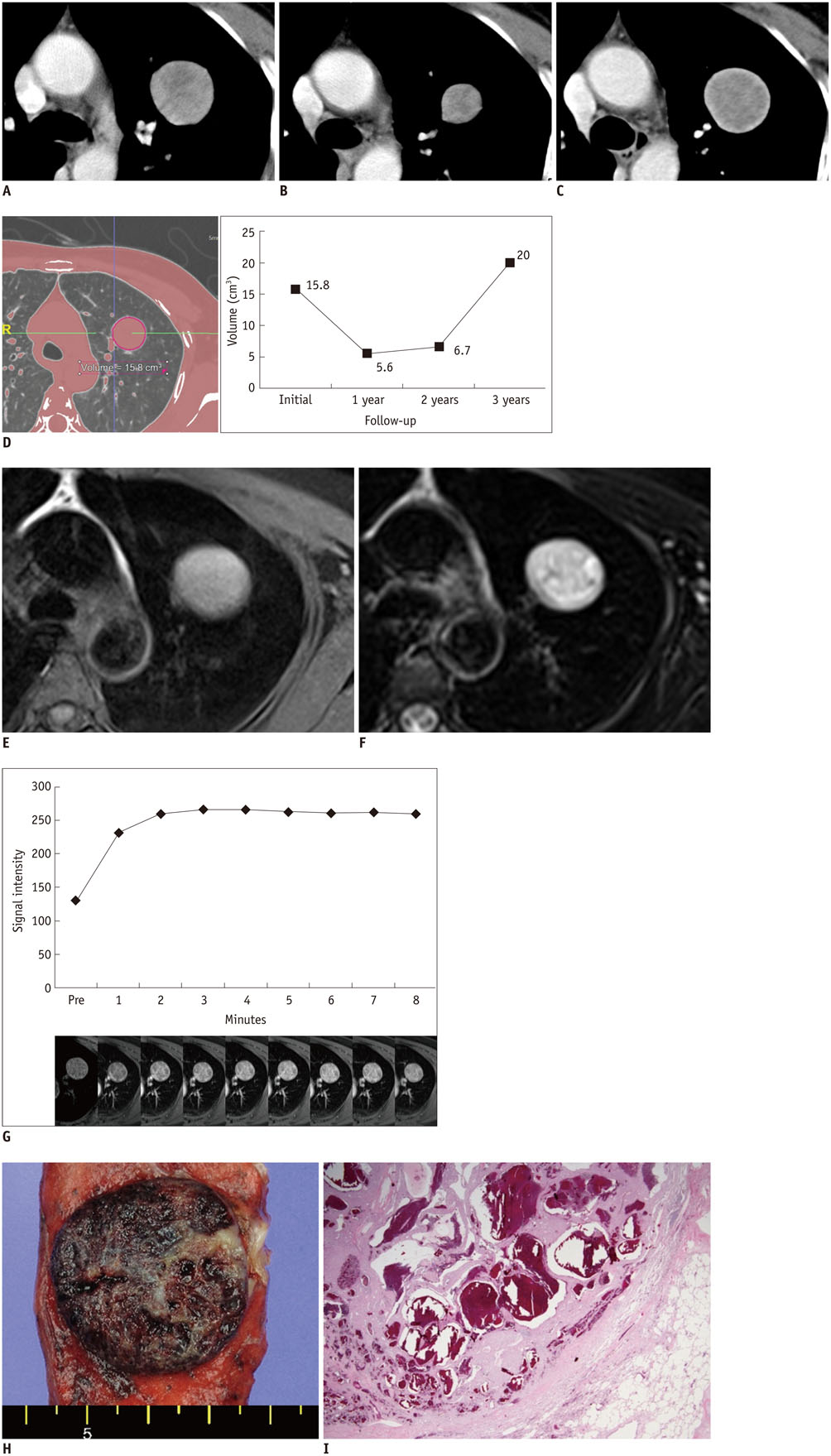
Fig. 1 Sclerosing pneumocytoma with wax-and-wane growth pattern in 47-year-old woman. A-C. Initial (A) and 1-year (B) and 3-year follow-up (C) contrast-enhanced computed tomography (CT) scans show heterogeneously enhancing mass in left upper lobe. Mass decreases in diameter from 3.1 to 2.1 cm and then marked increase to 3.4 cm on serial CT scans. D. Serial volume graph chart obtained from three-dimensional CT data using automated segmentation technique during follow-up shows wax-and-wane pattern. E, F. Mass shows iso- to higher signal intensity (SI) than that of muscle on T1-weighted magnetic resonance (MR) image (E) and heterogeneously high SI on fat-saturated T2-weighted image (F). G. Dynamic contrast-enhanced MR images and corresponding graph of SI versus time show early enhancement without peak point and subsequent plateau pattern. H. Gross findings show well-demarcated, solid mass with fibrous matrix and areas of hemorrhage. I. Well-demarcated mass with small cystic spaces filled with blood was observed on microscopic examination (hematoxylin and eosin staining, × 12.5).
Reference
-
1. Illei PB, Rosai J, Klimstra DS. Expression of thyroid transcription factor-1 and other markers in sclerosing hemangioma of the lung. Arch Pathol Lab Med. 2001; 125:1335–1339.2. Devouassoux-Shisheboran M, Hayashi T, Linnoila RI, Koss MN, Travis WD. A clinicopathologic study of 100 cases of pulmonary sclerosing hemangioma with immunohistochemical studies: TTF-1 is expressed in both round and surface cells, suggesting an origin from primitive respiratory epithelium. Am J Surg Pathol. 2000; 24:906–916.3. Fujiyoshi F, Ichinari N, Fukukura Y, Sasaki M, Hiraki Y, Nakajo M. Sclerosing hemangioma of the lung: MR findings and correlation with pathological features. J Comput Assist Tomogr. 1998; 22:1006–1008.4. Kim GY, Kim J, Choi YS, Kim HJ, Ahn G, Han J. Sixteen cases of sclerosing hemangioma of the lung including unusual presentations. J Korean Med Sci. 2004; 19:352–358.5. Liebow AA, Hubbell DS. Sclerosing hemangioma (histiocytoma, xanthoma) of the lung. Cancer. 1956; 9:53–75.6. Miyagawa-Hayashino A, Tazelaar HD, Langel DJ, Colby TV. Pulmonary sclerosing hemangioma with lymph node metastases: report of 4 cases. Arch Pathol Lab Med. 2003; 127:321–325.7. Kalhor N, Staerkel GA, Moran CA. So-called sclerosing hemangioma of lung: current concept. Ann Diagn Pathol. 2010; 14:60–67.8. Cheung YC, Ng SH, Chang JW, Tan CF, Huang SF, Yu CT. Histopathological and CT features of pulmonary sclerosing haemangiomas. Clin Radiol. 2003; 58:630–635.9. Im JG, Kim WH, Han MC, Han YM, Chung JW, Ahn JM, et al. Sclerosing hemangiomas of the lung and interlobar fissures: CT findings. J Comput Assist Tomogr. 1994; 18:34–38.10. Nakanishi K, Kohzaki S, Fujimoto S, Horita Y, Hayashi K. Pulmonary sclerosing hemangioma: report of a case with emphasis on dynamic MR imaging findings. Radiat Med. 1997; 15:117–119.11. Kono R, Fujimoto K, Terasaki H, Müller NL, Kato S, Sadohara J, et al. Dynamic MRI of solitary pulmonary nodules: comparison of enhancement patterns of malignant and benign small peripheral lung lesions. AJR Am J Roentgenol. 2007; 188:26–36.
- Full Text Links
-
- Actions
-
Cited
- CITED
-
- Close
- Share
-
- Similar articles
-
- Infected Sclerosing Lipogranuloma after Hernioplasty: Ultrasonographic and MRI Findings
- A study on the comparision of various imaging methods for the staging of renal cell carcinoma
- Retained Bone Wax on CT at One Year after Dacryocystorhinostomy: A Case Report
- A Case of Sclerosing Stromal Tumor of the Ovary
- CT and MRI Findings of Sclerosing Angiomatoid Nodular Transformation of the Spleen: Spoke Wheel Pattern
