Retained Bone Wax on CT at One Year after Dacryocystorhinostomy: A Case Report
- Affiliations
-
- 1Department of Radiology, Guri Hospital, Hanyang University College of Medicine, Guri, Korea. dwpark@hanyang.ac.kr
- 2Department of Radiology, Seoul Hospital, Hanyang University College of Medicine, Seoul, Korea.
- 3Department of Otolaryngology Head and Neck Surgery, Guri Hospital, Hanyang University College of Medicine, Guri, Korea.
- KMID: 2043611
- DOI: http://doi.org/10.3348/jksr.2015.73.3.190
Abstract
- A 71-year-old man with chronic rhinosinusitis presented with a purulent, foul-smelling nasal discharge and obstruction. One year earlier he had been treated with a dacryocystorhinostomy for nasolacrimal duct obstruction. During the procedure, bone wax had been used to control bleeding in the anterior upper nasal cavity. On computed tomographic imaging, a fat-density lesion was seen in the anterior upper sinonasal cavity and was found to be hypointense or signal-void on all magnetic resonance imaging sequences. The lesion, which proved to consist of bone wax, was surgically removed. Here, we present the imaging features of retained bone wax in a patient with clinically diagnosed chronic rhinosinusitis after dacryocystorhinostomy.
MeSH Terms
Figure
-

Fig. 1 Endoscopic view showing a whitish, glittering foreign body filling the anterior upper nasal cavity, medial to the upper attached part of the right middle turbinate.

Fig. 2 CT images obtained in a 71-year-old man with chronic sinusitis that developed one year after dacryocystorhinostomy. A. A pre-enhanced axial CT scan of the paranasal sinus shows a fat-density lesion (about -70 HU) filling the right anterior upper nasal cavity (arrow). B, C. Post-enhanced and coronal reformatted CT images in a bone window setting show a fat-density lesion (arrow) around the upper attached part of the middle turbinate and a right lacrimal bone defect adjacent to the lacrimal sac. The nasolacrimal duct remained patent (arrowhead) but was slightly compressed by the lesion. D. On axial CT scanning, a lump of bone wax (arrow) floating in a cup of water shows the low attenuation of fat (about -100 HU), as compared with the intermediate attenuation of water (about 0 HU). HU = Hounsfield units
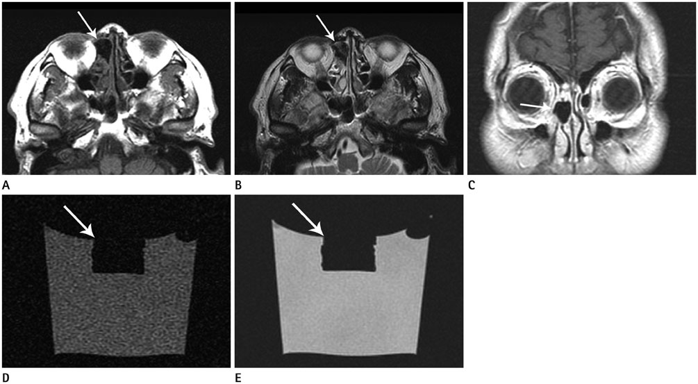
Fig. 3 After gadolinium injection, MRI scans were obtained showing a hypointense or signal-void lesion (arrow) on all sequences, including T1-weighted (A), T2-weighted (B), and axial and coronal contrast-enhanced T1-weighted (C) images. Signal-void coronal T1-weighted (D) and T2-weighted (E) MRI scans of a cube of bone wax floating in a cup of water.
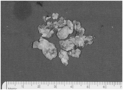
Fig. 4 Endoscopic sinus surgery was performed, and pieces of bone wax of various shapes and sizes (to a maximum of about 1.5 × 1.5 cm) were found and recovered.
Reference
-
1. Gupta G, Prestigiacomo CJ. From sealing wax to bone wax: predecessors to Horsley's development. Neurosurg Focus. 2007; 23:E16.2. Schonauer C, Tessitore E, Barbagallo G, Albanese V, Moraci A. The use of local agents: bone wax, gelatin, collagen, oxidized cellulose. Eur Spine J. 2004; 13:Suppl 1. S89–S96.3. Aurelio J, Chenail B, Gerstein H. Foreign-body reaction to bone wax. Report of a case. Oral Surg Oral Med Oral Pathol. 1984; 58:98–100.4. Hadeishi H, Yasui N, Suzuki A. Mastoid canal and migrated bone wax in the sigmoid sinus: technical report. Neurosurgery. 1995; 36:1220–1223. discussion 1223-12245. Katz SE, Rootman J. Adverse effects of bone wax in surgery of the orbit. Ophthal Plast Reconstr Surg. 1996; 12:121–126.6. Patel RB, Kwartler JA, Hodosh RM. Bone wax as a cause of foreign body granuloma in the cerebellopontine angle. Case illustration. J Neurosurg. 2000; 92:362.7. Kim KR, Cho SH, Jung JS, Choi SJ. A case of recurrent frontal sinusitis due to bone wax. J Rhinol. 2003; 10:64–67.8. Kamide T, Nakada M, Hirota Y, Hayashi Y, Hayashi Y, Uchiyama N, et al. Skull osteohypertrophy as a complication of bone wax. J Clin Neurosci. 2009; 16:1658–1660.9. Morrison PR, Silverman SG, Tuncali K, Tatli S. MRI-guided cryotherapy. J Magn Reson Imaging. 2008; 27:410–420.10. Allison RT. Foreign body reactions and an associated histological artefact due to bone wax. Br J Biomed Sci. 1994; 51:14–17.
- Full Text Links
-
- Actions
-
Cited
- CITED
-
- Close
- Share
-
- Similar articles
-
- Effect of the Bone wax for the Maintenance of a Created Fistula between the Maxillary Sinus and the Nasal Cavity in Rabbits
- A Case of Fungall Ball after External Dacryocystorhinostomy
- Application of Ultrasonic Bone Aspirator in Transnasal Endoscopic Dacryocystorhinostomy : A Case Report
- Dacryocystorhinostomy with Silicone Tube used in the Lacrimal Drainage System
- Benign Mass after Reduction Mandibular Angleplasty
