Korean J Radiol.
2008 Jul;9(Suppl):S52-S55. 10.3348/kjr.2008.9.s.s52.
CT and MRI Findings of Sclerosing Angiomatoid Nodular Transformation of the Spleen: Spoke Wheel Pattern
- Affiliations
-
- 1Department of Radiology, Hacettepe University School of Medicine, Ankara, Turkey. musturayk@yahoo.com
- KMID: 1100105
- DOI: http://doi.org/10.3348/kjr.2008.9.s.s52
Abstract
- Sclerosing angiomatoid nodular transformation of the spleen is a recently described benign pathologic entity that is characterized by round shaped vascular spaces that are lined by endothelial cells, and the spaces are circumscribed by granulomatoid structures. Microscopically, all the reported cases had multiple angiomatoid nodules in a fibrosclerotic stroma. Each angiomatoid nodule was made up of slit-like, round or irregular shaped vascular spaces that were lined by endothelial cells and interspersed ovoid cells. We present here the CT and dynamic gadolinium-enhanced MR findings of a patient with sclerosing angiomatoid nodular transformation. The spoke-wheel pattern that was observed on MRI in this case may be an important imaging clue for making the correct diagnosis of this benign lesion.
MeSH Terms
Figure
-
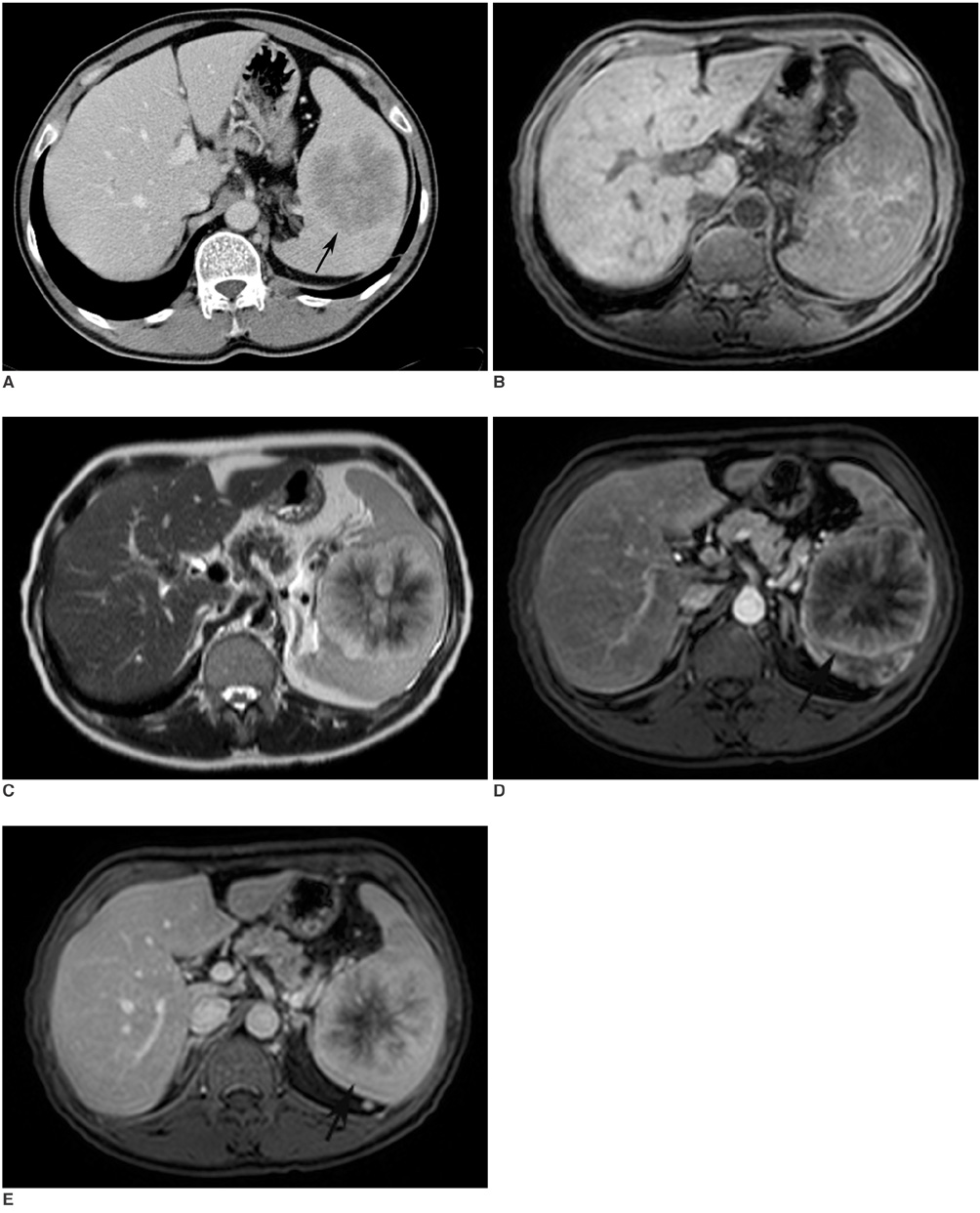
Fig. 1 Sclerosing angiomatoid nodular transformation in 44-year-old man. A. Axial CT image shows predominantly hypodense mass with lobulated contours. Also note rim-style contrast enhancement of external borders of lesion (arrow). B. Fat-saturated precontrast T1-weighted image shows central hyperintensity that is consistent with hemorrhage. C. T2-weighted MR image shows spoke-wheel pattern of lesion that is predominantly hyperintense with central hypointense areas. Note hyperintense radiations towards center of lesion. D, E Postcontrast arterial (D) and delayed venous phase (E) T1 weighted MR images clearly show progressive enhancement from periphery to center of lesion, which is similar to spoke wheel pattern (arrows).
Reference
-
1. Elsayes KM, Narra VR, Mukundan G, Lewis JS Jr, Menias CO, Heiken JP. MR imaging of the spleen: spectrum of abnormalities. Radiographics. 2005. 25:967–982.2. Martel M, Cheuk W, Lombardi L, Lifschitz-Mercer B, Chan JK, Rosai J. Sclerosing angiomatoid nodular transformation (SANT): report of 25 cases of a distinctive benign splenic lesion. Am J Surg Pathol. 2004. 28:1268–1279.3. Li L, Fisher DA, Stanek AE. Sclerosing angiomatoid nodular transformation (SANT) of the spleen: addition of a case with focal CD68 staining and distinctive CT features. Am J Surg Pathol. 2005. 29:839–841.4. Rosai J. Rosai and Ackerman's surgical pathology. 2004. 9th ed. Edinburgh: Mosby;2035.
- Full Text Links
-
- Actions
-
Cited
- CITED
-
- Close
- Share
-
- Similar articles
-
- Splenic Sclerosing Angiomatoid Nodular Transformation in an 8-Year-Old Child
- A Case of Sclerosing Angiomatoid Nodular Transformation of the Spleen: Spoke Wheel Pattern on Computed Tomography
- Laparoscopic splenectomy for sclerosing angiomatoid nodular transformation of the spleen
- Sclerosing Angiomatoid Nodular Transformation (SANT) in Spleen: A Case Report
- Multimodality Imaging Features of Various Splenic Lesions: Clinical and Histopathologic Correlation
