Should We Recommend Ultrasonography for an Incidental Thyroid Nodule on Additional Cervicothoracic Sagittal T2-Weighted Image of Lumbar Spine MRI?
- Affiliations
-
- 1Department of Radiology, Research Institute of Radiological Science, Medical Convergence Research Institute, and Severance Biomedical Science Institute, Yonsei University College of Medicine, Seoul, Korea.
- 2Department of Orthopedic Surgery, Yonsei University College of Medicine, Seoul, Korea. parkjo@yuhs.ac
- KMID: 2151769
- DOI: http://doi.org/10.13104/imri.2015.19.4.224
Abstract
- PURPOSE
To determine whether we should recommend ultrasonography (US) for an incidental thyroid nodule identified by additional cervicothoracic sagittal T2-weighted image (C-T sag T2WI) of lumbar spine magnetic resonance imaging (MRI).
MATERIALS AND METHODS
A retrospective study of 61 patients who underwent both lumbar spine MRI and thyroid US between December 2011 and April 2015 was conducted. For all US-found thyroid nodules > 1 cm, investigators evaluated whether there was any correlation between thyroid nodule detectability by C-T sag T2WI and US features such as echogenicity, composition, or suspicion of malignancy.
RESULTS
Solid hypoechoic (2/4; 50%) or mixed echoic nodules (4/8; 50%) appeared to be found relatively more easily by C-T sag T2WI than more benign-looking solid isoechoic (1/4; 25%) or spongiform nodules (0/6; 0%). Among six nodules with ultrasonographic suspicion for malignancy, only one nodule was detected by C-T sag T2WI.
CONCLUSION
If an incidental thyroid nodule is seen by C-T sag T2WI, it would be better to recommend thyroid US for identifying malignancy.
MeSH Terms
Figure
-
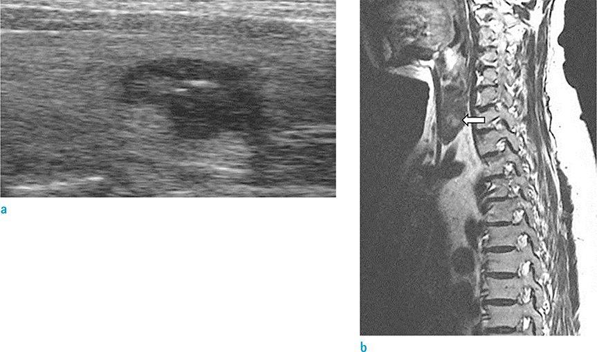
Fig. 1 A 65-year-old woman with a 12-mm right thyroid nodule. (a) Thyroid US reveals an unparalleled solid hypoechoic thyroid nodule with microcalcifications, which is suspicious for malignancy. The nodule was diagnosed as benign by fine needle aspiration biopsy. (b) After 3 months, C-T sag T2WI of lumbar spine MRI for evaluation of lower back pain shows a T2-high signal intensity nodule in the thyroid (arrow).
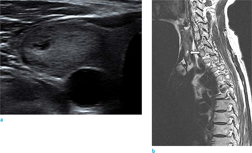
Fig. 2 A 74-year-old woman with a 25-mm right thyroid nodule. (a) The thyroid nodule shows mixed solid and cystic composition in the US. (b) C-T sag T2WI of lumbar spine MRI two days later shows a nodule with cystic component in the thyroid (arrow). It can be mistaken for a much smaller nodule in the MR images.

Fig. 3 Undetected > 1-cm thyroid nodules. (a, b) A spongiform thyroid nodule of a 55-year-old woman. (c, d) A solid isoechoic thyroid nodule of a 56-year-old woman. These nodules were not identified by C-T sag T2WI of lumbar spine MRI.
Reference
-
1. Kamath S, Jain N, Goyal N, Mansour R, Mukherjee K. Incidental findings on MRI of the spine. Clin Radiol. 2009; 64:353–361.2. Grady AT, Sosa JA, Tanpitukpongse TP, Choudhury KR, Gupta RT, Hoang JK. Radiology reports for incidental thyroid nodules on CT and MRI: high variability across subspecialties. AJNR Am J Neuroradiol. 2015; 36:397–402.3. Nam-Goong IS, Kim HY, Gong G, et al. Ultrasonography-guided fine-needle aspiration of thyroid incidentaloma: correlation with pathological findings. Clin Endocrinol (Oxf). 2004; 60:21–28.4. Kang HW, No JH, Chung JH, et al. Prevalence, clinical and ultrasonographic characteristics of thyroid incidentalomas. Thyroid. 2004; 14:29–33.5. Kim K, Emoto N, Mishina M, et al. Incidental detection of thyroid nodules at magnetic resonance imaging of the cervical spine. Neurol Med Chir (Tokyo). 2013; 53:77–81.6. Seo J, Park SY, Lee JW, Lee GY, Kang HS. The value of additional cervicothoracic spine sagittal T2-weighted images included in routine lumbar spine MR imaging. J Korean Soc Magn Reson Med. 2013; 17:91–100.7. Cooper DS, Doherty GM, Haugen BR, et al. Revised American Thyroid Association management guidelines for patients with thyroid nodules and differentiated thyroid cancer: the American Thyroid Association (ATA) guidelines taskforce on thyroid nodules and differentiated thyroid cancer. Thyroid. 2009; 19:1167–1214.8. Kwak JY, Han KH, Yoon JH, et al. Thyroid imaging reporting and data system for US features of nodules: a step in establishing better stratification of cancer risk. Radiology. 2011; 260:892–899.9. Serpell J. Management guidelines for patients with thyroid nodules. ANZ J Surg. 2010; 80:765–766.10. Cooper DS, Doherty GM, Haugen BR, et al. Management guidelines for patients with thyroid nodules and differentiated thyroid cancer. Thyroid. 2006; 16:109–142.11. Kamran SC, Marqusee E, Kim MI, et al. Thyroid nodule size and prediction of cancer. J Clin Endocrinol Metab. 2013; 98:564–570.12. Wong KT, Ahuja AT. Ultrasound of thyroid cancer. Cancer Imaging. 2005; 5:157–166.13. Moon WJ, Jung SL, Lee JH, et al. Benign and malignant thyroid nodules: US differentiation--multicenter retrospective study. Radiology. 2008; 247:762–770.14. Lee MJ, Kim EK, Kwak JY, Kim MJ. Partially cystic thyroid nodules on ultrasound: probability of malignancy and sonographic differentiation. Thyroid. 2009; 19:341–346.15. Bellantone R, Lombardi CP, Raffaelli M, et al. Management of cystic or predominantly cystic thyroid nodules: the role of ultrasound-guided fine-needle aspiration biopsy. Thyroid. 2004; 14:43–47.
- Full Text Links
-
- Actions
-
Cited
- CITED
-
- Close
- Share
-
- Similar articles
-
- The Value of Additional Cervicothoracic Spine Sagittal T2-weighted Images Included in Routine Lumbar Spine MR Imaging
- Incorporation of Whole Spine Screening in Magnetic Resonance Imaging Protocols for Low Back Pain: A Valuable Addition
- Clinical Utility of Limited T2-Weighted-Only Lumbar Spine MRI in Pain Intervention Clinics
- Value of Additional Cervicothoracic Sagittal T2-Weighted Images in Elderly Patients with Symptoms Suggestive of Lumbar Spinal Stenosis
- Coexisting Spine Lesions on Whole Spine T2 Sagittal MRI in Evaluating Spinal Degenerative Disease
