The Value of Additional Cervicothoracic Spine Sagittal T2-weighted Images Included in Routine Lumbar Spine MR Imaging
- Affiliations
-
- 1Department of Radiology, Seoul National University Bundang Hospital, Seongnam-si, Gyeonggi-do, Korea. joonwoo2@gmail.com
- KMID: 2099868
- DOI: http://doi.org/10.13104/jksmrm.2013.17.2.91
Abstract
- PURPOSE
To evaluate the usefulness of cervicothoracic spine sagittal T2-weighted images (CT SAG T2WIs) included in routine lumbar spine MRI.
MATERIALS AND METHODS
Institutional review board approval was obtained and informed consents were waived for this retrospective study. The study group comprised 2,113 patients who underwent lumbar spine MRI from January 2005 to December 2005. CT SAG T2WIs were added in the routine lumbar spine MRIs. Radiologic reports were reviewed retrospectively for pathologic lesions on CT SAG T2WIs by one radiologist. Information of additional cervical or thoracic spine MRI and/or CT for further evaluation of positive findings on CT SAG T2WIs and their treatment were collected by retrospectively reviewing medical records.
RESULTS
The CT SAG T2WIs revealed 142 pathologic lesions in 139 (6.58%) of the 2,113 patients. They were easily obtained without positional change in a scan time of less than 2 minutes. Additional cervical or thoracic spine MRI and/or CT for positive findings on CT SAG T2WIs were performed in 13 patients. Seven patients underwent surgical treatment.
CONCLUSION
CT SAG T2WIs included in routine lumbar spine MRI were useful in finding the pathologic lesions in cervicothoracic spine for the patients who assumed to have lesions in lumbar spine.
Figure
-
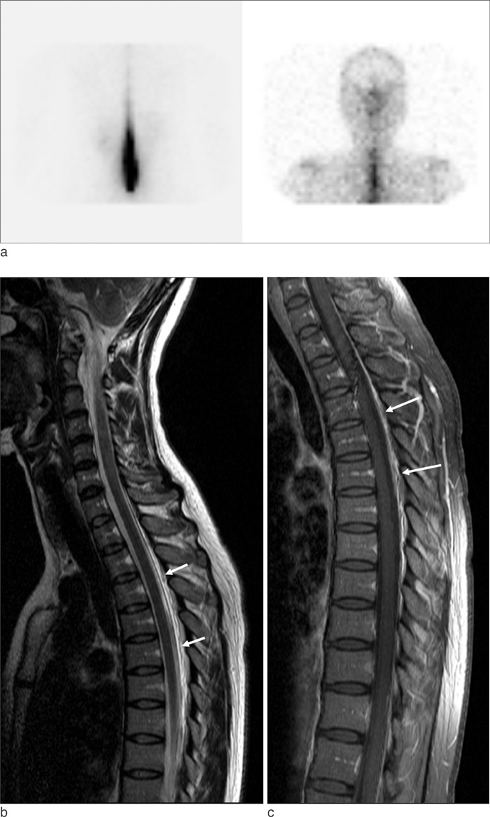
Fig. 1 33-year-old woman with headache due to spontaneous intracranial hypotension. a. Six-hour delayed image of cisternography shows remaining radiotracer at lumbar spine level and delayed migration. b. Additional cervicothoracic sagittal T2-weighted image included in lumbar spine MRI demonstrates posterior epidural fluid (arrows) at thoracic spine level. c. Gadolinium-enhanced fat-suppressed sagittal T1-weighted image performed 8 hours later shows enhancement of posterior epidural space (arrows).
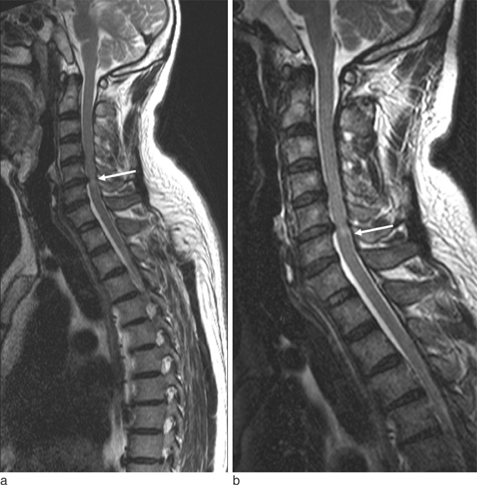
Fig. 2 50-year-old man with paraplegia due to compressive myelopathy associated with cervical disc herniation. a. Cervicothoracic sagittal T2-weighted image included in lumbar spine MRI shows herniated disc and intramedullary high signal intensity (arrow) at the level of C5-6 disc. b. Cervical spine MRI was performed one day later. Sagittal T2-weighted image demonstrates disc herniation and intramedullary high signal intensity at C5-6 level.
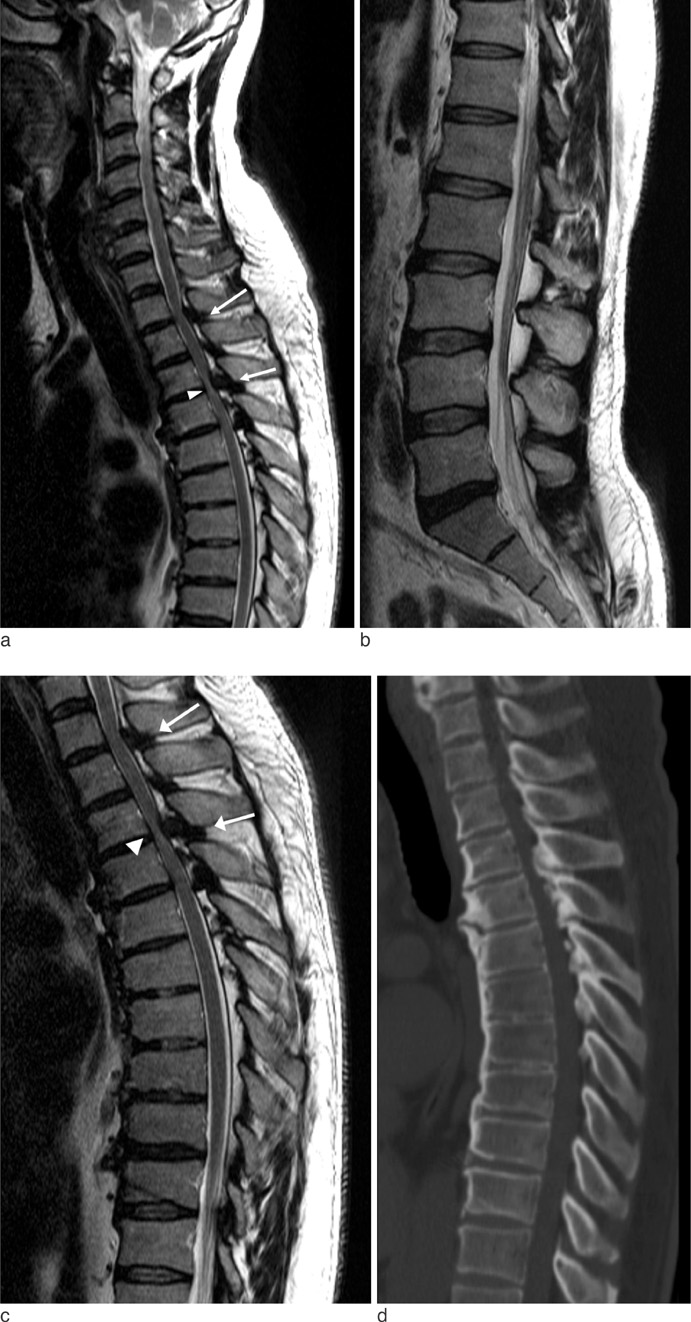
Fig. 3 52-year-old man with lower back pain, radiating pain, and numbness at both medial side of thigh, anterior lower legs, and feet due to spinal canal stenosis associated with ossification of ligamentum flavum. a. Cervicothoracic sagittal T2-weighted image included in lumbar spine MRI shows hypertrophy of the ligamentum flavum at the T1-2 and T3-4 levels (arrows), and suspicious intramedullary high SI at T3-4 levels (white arrowhead). b. Sagittal T2-weighted image of the lumbar spine shows mild disc bulging and degeneration at L5-S1. c, d. Thoracic spine MRI and CT were performed three days later. Sagittal T2-weighted image (c) demonstrates hypertrophy of the ligamenta flava at the T1-2 and T3-4 levels (arrows) and consistent intramedullary high signal intensity at the T3-4 level (white arrowhead). Sagittal reformatted CT image (d) shows ossification of the ligamenta flava with spinal canal narrowing at the T3-4 level.

Fig. 4 46-year-old woman with radiating pain in right leg due to idiopathic spinal cord herniation. a. Cervicothoracic sagittal T2-weighted image included in lumbar spine MR imaging shows a focal anterior kink of the spinal cord at the level of T3-4 (arrowhead). b, c. Thoracic spine MRI was performed 23 days later. Sagittal T2-weighted (b) and axial T1-weighted (c) images clearly demonstrate herniation of the left anterolateral portion of the cord through a dural defect. d. Sagittal T2-weighted image in thoracic spine MR imaging performed three years later shows reduction of spinal cord herniation and suspicious anterior subdural or epidural fluid collection at thoracic and lumbar spine level.
Reference
-
1. Muhle C, Metzner J, Weinert D, et al. Classification system based on kinematic MR imaging in cervical spondylitic myelopathy. AJNR Am J Neuroradiol. 1998; 19:1763–1771.2. Potter K, Saifuddin A. MRI of chronic spinal cord injury. British Journal of Radiology. 2003; 76:347–352.3. Watters MR, Stears JC, Osborn AG, et al. Transdural spinal cord herniation: imaging and clinical spectra. AJNR Am J Neuroradiol. 1998; 19:1337–1344.4. Chen CJ, Lee TH, Hsu HL, Tseng YC, Wong YC, Wang LJ. Spinal MR findings in spontaneous intracranial hypotension. Neuroradiology. 2002; 44:996–1003.5. LaBan MM, Green ML. Concurrent (tandem) cervical and lumbar spinal stenosis: a 10-yr review of 54 hospitalized patients. Am J Phys Med Rehabil. 2004; 83:187–190.6. Lee MJ, Garcia R, Cassinelli EH, Furey C, Riew KD. Tandem stenosis: a cadaveric study in osseous morphology. The Spine Journal. 2008; 8:1003–1006.7. Dagi TF, Tarkington MA, Leech JJ. Tandem lumbar and cervical spinal stenosis. Natural history, prognostic indices, and results after surgical decompression. J Neurosurg. 1987; 66:842–849.8. Edwards CC 2nd, Riew KD, Anderson PA, Hilibrand AS, Vaccaro AF. Cervical myelopathy. current diagnostic and treatment strategies. Spine J. 2003; 3:68–81.9. Slipman CW, Shin CH, Patel RK, et al. Etiologies of failed back surgery syndrome. Pain Med. 2002; 3:200–214. discussion 214-207.10. Parmar H, Park P, Brahma B, Gandhi D. Imaging of idiopathic spinal cord herniation. Radiographics. 2008; 28:511–518.11. Wada E, Yonenobu K, Kang J. Idiopathic spinal cord herniation: report of three cases and review of the literature. Spine (Phila Pa 1976). 2000; 25:1984–1988.12. Gandhi D, Goyal M, Bourque PR. Case 138: Idiopathic spinal cord herniation. Radiology. 2008; 249:384–388.
- Full Text Links
-
- Actions
-
Cited
- CITED
-
- Close
- Share
-
- Similar articles
-
- Should We Recommend Ultrasonography for an Incidental Thyroid Nodule on Additional Cervicothoracic Sagittal T2-Weighted Image of Lumbar Spine MRI?
- Value of Additional Cervicothoracic Sagittal T2-Weighted Images in Elderly Patients with Symptoms Suggestive of Lumbar Spinal Stenosis
- Clinical Utility of Limited T2-Weighted-Only Lumbar Spine MRI in Pain Intervention Clinics
- Coexisting Spine Lesions on Whole Spine T2 Sagittal MRI in Evaluating Spinal Degenerative Disease
- Incorporation of Whole Spine Screening in Magnetic Resonance Imaging Protocols for Low Back Pain: A Valuable Addition
