Immunostimulatory Effects of Silica Nanoparticles in Human Monocytes
- Affiliations
-
- 1Department of Microbiology, The Institute for Immunology and Immunological Diseases, Yonsei University College of Medicine, Seoul 120-752, Korea. inhong@yuhs.ac
- KMID: 2150772
- DOI: http://doi.org/10.4110/in.2013.13.3.94
Abstract
- Amorphous silica particles, whose applications are increasing in many biomedical fields, are known to be less toxic than crystalline silica. In this study, the inflammatory effects of amorphous silica nanoparticles were investigated using 30-nm amorphous silica nanoparticles and human peripheral blood mononuclear cells (PBMCs) or purified monocytes. As a result, production of IL-1beta and IL-8 were increased. In addition, the mitochondrial reactive oxygen species (ROS) was detected, which may lead to mitochondrial membrane disruption. Most importantly, inflammasome formation was observed. Therefore, these results provide immunological information about amorphous silica nanoparticles and suggest that amorphous silica nanoparticles can evoke innate immune reactions in human monocytes through production of IL-1beta and IL-8.
MeSH Terms
Figure
-

Figure 1 Cytotoxicity and production of cytokines in PBMCs. (a) PBMCs were treated with 30-nm amorphous silica nanoparticles for 6 h and cytotoxicity was determined by CCK-8 assay. (b, c) PBMCs were treated with silica nanoparticles for 6 h and supernatant levels of IL-1β (b) and IL-8 (c) were assessed by ELISA. LPS (25 ng/ml) was pretreated for 3 h before nanoparticle exposure. Data represent means±S.D. of three independent experiments. One-way ANOVA analysis shows significance (p<0.0001) (b, c) and student's t-test between certain pairs (b, c) was used for statistical analysis.

Figure 2 Production of mitochondria ROS in blood monocytes. Monocytes were treated with silica nanoparticles or silver nanoparticles for 40 min. Cells were stained with 2.5µM MitoSOX (for mitochondrial superoxide) for 15 min and analyzed by flow cytometry. MI, mean intensity. Data shown here are representative results from three independent experiments.
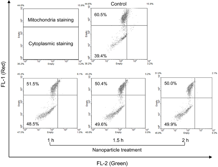
Figure 3 Assessment of mitochondrial membrane integrity. Monocytes were treated with 125µg/ml of 30 nm silica nanoparticles for 1 h, 1.5 h, and 2 h. After exposure for the indicated time, JC-1 (2µM) staining was performed to evaluate disturbance of mitochondrial membranes. Cells stained as red (intact mitochondria) were determined by flow cytometry. Data shown here are representative results from three independent experiments. MI, mean intensity.
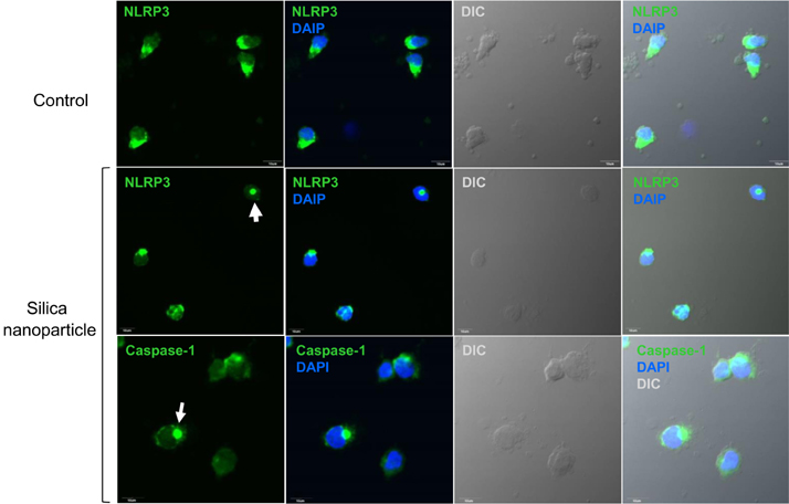
Figure 4 Inflammasome formation of caspase-1 and NLRP3 in monocytes. To analyze oligomerization of NLRP3 and caspase-1, monocytes were treated with silica nanoparticles (125µg/ml) for 15 min. Oligomerization of NLRP3 and caspase-1 was assessed by confocal microscopy after staining with anti-NLRP3 and anti-caspase-1 antibodies. White arrows indicate oligomerization of each molecule, which are not observed in controls.
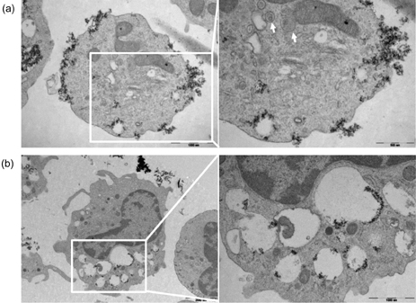
Figure 5 TEM images of monocytes after exposure to silica nanoparticles. Monocytes were treated with silica nanoparticles (125µg/ml) for 30 min. After 30 nm silica nanoparticle exposure, (a) double membrane structures (white arrows) suggesting autophagy-like structures and (b) presence of silica nanoparticles were observed inside endosome.
Reference
-
1. Hirsch LR, Stafford RJ, Bankson JA, Sershen SR, Rivera B, Price RE, Hazle JD, Halas NJ, West JL. Nanoshell-mediated near-infrared thermal therapy of tumors under magnetic resonance guidance. Proc Natl Acad Sci U S A. 2003; 100:13549–13554.
Article2. Bharali DJ, Klejbor I, Stachowiak EK, Dutta P, Roy I, Kaur N, Bergey EJ, Prasad PN, Stachowiak MK. Organically modified silica nanoparticles: a nonviral vector for in vivo gene delivery and expression in the brain. Proc Natl Acad Sci U S A. 2005; 102:11539–11544.
Article3. Roy I, Ohulchanskyy TY, Bharali DJ, Pudavar HE, Mistretta RA, Kaur N, Prasad PN. Optical tracking of organically modified silica nanoparticles as DNA carriers: a nonviral, nanomedicine approach for gene delivery. Proc Natl Acad Sci U S A. 2005; 102:279–284.
Article4. Bottini M, D'Annibale F, Magrini A, Cerignoli F, Arimura Y, Dawson MI, Bergamaschi E, Rosato N, Bergamaschi A, Mustelin T. Quantum dot-doped silica nanoparticles as probes for targeting of T-lymphocytes. Int J Nanomedicine. 2007; 2:227–233.5. Verraedt E, Pendela M, Adams E, Hoogmartens J, Martens JA. Controlled release of chlorhexidine from amorphous microporous silica. J Control Release. 2010; 142:47–52.
Article6. Zhang FF, Wan Q, Li CX, Wang XL, Zhu ZQ, Xian YZ, Jin LT, Yamamoto K. Simultaneous assay of glucose, lactate, L-glutamate and hypoxanthine levels in a rat striatum using enzyme electrodes based on neutral red-doped silica nanoparticles. Anal Bioanal Chem. 2004; 380:637–642.
Article7. Santra S, Zhang P, Wang K, Tapec R, Tan W. Conjugation of biomolecules with luminophore-doped silica nanoparticles for photostable biomarkers. Anal Chem. 2001; 73:4988–4993.
Article8. Gemeinhart RA, Luo D, Saltzman WM. Cellular fate of a modular DNA delivery system mediated by silica nanoparticles. Biotechnol Prog. 2005; 21:532–537.
Article9. Slowing II, Vivero-Escoto JL, Wu CW, Lin VS. Mesoporous silica nanoparticles as controlled release drug delivery and gene transfection carriers. Adv Drug Deliv Rev. 2008; 60:1278–1288.
Article10. Maynard AD, Aitken RJ, Butz T, Colvin V, Donaldson K, Oberdörster G, Philbert MA, Ryan J, Seaton A, Stone V, Tinkle SS, Tran L, Walker NJ, Warheit CB. Safe handling of nanotechnology. Nature. 2006; 444:267–269.
Article11. Greenberg MI, Waksman J, Curtis J. Silicosis: a review. Dis Mon. 2007; 53:394–416.
Article12. Mossman BT, Churg A. Mechanisms in the pathogenesis of asbestosis and silicosis. Am J Respir Crit Care Med. 1998; 157:1666–1680.
Article13. Huaux F. New developments in the understanding of immunology in silicosis. Curr Opin Allergy Clin Immunol. 2007; 7:168–173.
Article14. Yang EJ, Kim S, Kim JS, Choi IH. Inflammasome formation and IL-1β release by human blood monocytes in response to silver nanoparticles. Biomaterials. 2012; 33:6858–6867.
Article15. Hornung V, Bauernfeind F, Halle A, Samstad EO, Kono H, Rock KL, Fitzgerald KA, Latz E. Silica crystals and aluminum salts activate the NALP3 inflammasome through phagosomal destabilization. Nat Immunol. 2008; 9:847–856.
Article16. Dostert C, Pétrilli V, Van Bruggen R, Steele C, Mossman BT, Tschopp J. Innate immune activation through Nalp3 inflammasome sensing of asbestos and silica. Science. 2008; 320:674–677.
Article17. Cassel SL, Eisenbarth SC, Iyer SS, Sadler JJ, Colegio OR, Tephly LA, Carter AB, Rothman PB, Flavell RA, Sutterwala FS. The Nalp3 inflammasome is essential for the development of silicosis. Proc Natl Acad Sci U S A. 2008; 105:9035–9040.
Article18. Morishige T, Yoshioka Y, Inakura H, Tanabe A, Yao X, Narimatsu S, Monobe Y, Imazawa T, Tsunoda S, Tsutsumi Y, Mukai Y, Okada N, Nakagawa S. The effect of surface modification of amorphous silica particles on NLRP3 inflammasome mediated IL-1beta production, ROS production and endosomal rupture. Biomaterials. 2010; 31:6833–6842.
Article19. Winter M, Beer HD, Hornung V, Krämer U, Schins RP, Förster I. Activation of the inflammasome by amorphous silica and TiO2 nanoparticles in murine dendritic cells. Nanotoxicology. 2011; 5:326–340.
Article20. Yazdi AS, Guarda G, Riteau N, Drexler SK, Tardivel A, Couillin I, Tschopp J. Nanoparticles activate the NLR pyrin domain containing 3 (Nlrp3) inflammasome and cause pulmonary inflammation through release of IL-1α and IL-1β. Proc Natl Acad Sci U S A. 2010; 107:19449–19454.
Article21. Jin C, Flavell RA. Molecular mechanism of NLRP3 inflammasome activation. J Clin Immunol. 2010; 30:628–631.
Article22. Netea MG, Nold-Petry CA, Nold MF, Joosten LA, Opitz B, van der Meer JH, van de Veerdonk FL, Ferwerda G, Heinhuis B, Devesa I, Funk CJ, Mason RJ, Kullberg BJ, Rubartelli A, van der Meer JW, Dinarello CA. Differential requirement for the activation of the inflammasome for processing and release of IL-1beta in monocytes and macrophages. Blood. 2009; 113:2324–2335.
Article23. Landmesser U, Dikalov S, Price SR, McCann L, Fukai T, Holland SM, Mitch WE, Harrison DG. Oxidation of tetrahydrobiopterin leads to uncoupling of endothelial cell nitric oxide synthase in hypertension. J Clin Invest. 2003; 111:1201–1209.
Article24. Sevier CS, Kaiser CA. Ero1 and redox homeostasis in the endoplasmic reticulum. Biochim Biophys Acta. 2008; 1783:549–556.
Article25. Sorbara MT, Girardin SE. Mitochondrial ROS fuel the inflammasome. Cell Res. 2011; 21:558–560.
Article26. Shukla RK, Kumar A, Pandey AK, Singh SS, Dhawan A. Titanium dioxide nanoparticles induce oxidative stress-mediated apoptosis in human keratinocyte cells. J Biomed Nanotechnol. 2011; 7:100–101.
Article27. Ahmad J, Ahamed M, Akhtar MJ, Alrokayan SA, Siddiqui MA, Musarrat J, Al-Khedhairy AA. Apoptosis induction by silica nanoparticles mediated through reactive oxygen species in human liver cell line HepG2. Toxicol Appl Pharmacol. 2012; 259:160–168.
Article28. Bulua AC, Simon A, Maddipati R, Pelletier M, Park H, Kim KY, Sack MN, Kastner DL, Siegel RW. Mitochondrial reactive oxygen species promote production of proinflammatory cytokines and are elevated in TNFR1-associated periodic syndrome (TRAPS). J Exp Med. 2011; 208:519–533.
Article29. Arnoult D, Soares F, Tattoli I, Girardin SE. Mitochondria in innate immunity. EMBO Rep. 2011; 12:901–910.
Article
- Full Text Links
-
- Actions
-
Cited
- CITED
-
- Close
- Share
-
- Similar articles
-
- The Effects of Silica Nanoparticles in Macrophage Cells
- Induction of Functional Changes of Dendritic Cells by Silica Nanoparticles
- Silica-Capped and Gold-Decorated Silica Nanoparticles for Enhancing Effect of Gold Nanoparticle-Based Photothermal Therapy
- Silica-Based Advanced Nanoparticles For Treating Ischemic Disease
- Color alterations of a PMMA resin for fixed interim prostheses reinforced with silica nanoparticles
