A Patient With Focal Dystonia That Occurred Secondary to a Peripheral Neurogenic Tumor: A Case Report
- Affiliations
-
- 1Department of Physical Medicine and Rehabilitation, Kyung Hee University College of Medicine, Seoul, Korea. kimhsmd@khu.ac.kr
- KMID: 2148240
- DOI: http://doi.org/10.5535/arm.2015.39.4.654
Abstract
- Dystonia is a movement disorder characterized by involuntary muscle contractions. Patients with dystonia may experience uncontrollable twisting, repetitive movements, or abnormal posture. A 55-year-old man presented with an involuntary left forearm supination, which he had experienced for five years. There was no history of antecedent trauma to the wrist or elbow. Although conventional therapeutic modalities had been performed, the symptoms persisted. When he visited our hospital, electromyography was performed. Reduced conduction velocity was evident at the elbow-axilla segment of the left median nerve. We suspected that there was a problem on the median nerve between the elbow and the axilla. For this reason, we performed an ultrasonography and magnetic resonance imaging study. A spindle-shaped soft tissue mass was observed at the left median nerve that suggested the possibility of neurofibroma. Dystonia caused by traumatic or compressive peripheral nerve injury has often been reported, but focal dystonia due to a neurogenic tumor is extremely rare. Here, we report our case with a review of the literature.
Keyword
MeSH Terms
Figure
-

Fig. 1 Clinical presentation. (A) Involuntary forearm supination with elbow flexed at 90°. (B) He had to type on a keyboard by raising his elbow due to forearm supination.

Fig. 2 Cervical magnetic resonance image taken at another hospital. (A) Sagittal image demonstrates disc space narrowing at C5-6 and multi-level disc protrusion. (B) Axial image demonstrates right paracentral disc protrusion at C5-6 level.
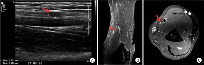
Fig. 3 Imaging study of left upper extremity. (A) Longitudinal ultrasonographic scan of distal arm demonstrates nodular shaped mass in the median nerve. (B) T1 sagittal image with gadolinium enhancement demonstrates a high signal nodular lesion at the distal arm. (C) T1 transverse image with gadolinium enhancement demonstrates a high signal target appearance mass in the median nerve at the distal arm.
Reference
-
1. Albanese A, Bhatia K, Bressman SB, Delong MR, Fahn S, Fung VS, et al. Phenomenology and classification of dystonia: a consensus update. Mov Disord. 2013; 28:863–873. PMID: 23649720.
Article2. Jankovic J. Peripherally induced movement disorders. Neurol Clin. 2009; 27:821–832. PMID: 19555833.
Article3. Iyer V, Thirkannad S. Focal hand dystonia in a patient with ulnar nerve neuropathy at the elbow. Hand. 2010; 5:453–457. PMID: 22131933.
Article4. Quartarone A, Morgante F, Sant'angelo A, Rizzo V, Bagnato S, Terranova C, et al. Abnormal plasticity of sensorimotor circuits extends beyond the affected body part in focal dystonia. J Neurol Neurosurg Psychiatry. 2008; 79:985–990. PMID: 17634214.
Article
- Full Text Links
-
- Actions
-
Cited
- CITED
-
- Close
- Share
-
- Similar articles
-
- Botulinum Toxin Injection Therapy for Lingual Dystonia: A Case Report
- Delayed-onset focal dystonia after stroke
- Delayed-onset focal dystonia after diffuse cerebral hypoxia: two case reports
- Unusual focal dyskinesia: the ears and abdomen
- Fibromyalgia Complicated with Dystonia Successfully Treated with Deep Brain Stimulation: a Case Report and Review of the Literature
