Clin Endosc.
2012 Sep;45(3):269-273.
Role of Computed Tomography Enterography/Magnetic Resonance Enterography: Is It in Prime Time?
- Affiliations
-
- 1Department of Radiology and Research Institute of Radiology, Asan Medical Center, University of Ulsan College of Medicine, Seoul, Korea. aykim@amc.seoul.kr
Abstract
- Today, cross-sectional imaging modalities, such as computed tomography enterography (CTE) and magnetic resonance enterography (MRE), are particularly suited to evaluate small bowel diseases, especially Crohn's disease (CD). It is well known that CTE/MRE can provide excellent assessment of disease activity as well as the macroscopic features, extramural abnormalities, and complications of the small intestine in patients with CD. In general, CTE is considered as the first-line modality for the evaluation of suspected inflammatory bowel disease and for long-term assessment or follow-up of these patients. Because of the advantage of lack of radiation, MRE is being used more frequently, especially in children or young patients with CD.
Keyword
MeSH Terms
Figure
-
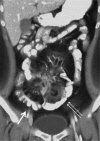
Fig. 1 Computed tomography enterography (CTE) of 26-year-old male with active Crohn's disease. On coronal volume rendering image of CTE shows increased attenuation in perienteric fat (double arrows), mesenteric haziness, and engorged vasa recta (thick arrow) along small intestine, indicating active inflammation, with enteroenteric fistulous tracts.
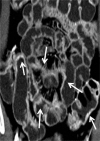
Fig. 2 Magnetic resonance enterography (MRE) of 37-year-old male with active Crohn's disease. Coronal reconstruction image of arterial-phase MRE demonstrates multifocal eccentric bowel wall thickening with intense mural enhancement of small intestine (arrows), indicating active inflammation.
Reference
-
1. Tochetto S, Yaghmai V. CT enterography: concept, technique, and interpretation. Radiol Clin North Am. 2009; 47:117–132. PMID: 19195538.
Article2. Huprich JE, Fletcher JG. CT enterography: principles, technique and utility in Crohn's disease. Eur J Radiol. 2009; 69:393–397. PMID: 19118968.
Article3. Paulsen SR, Huprich JE, Fletcher JG, et al. CT enterography as a diagnostic tool in evaluating small bowel disorders: review of clinical experience with over 700 cases. Radiographics. 2006; 26:641–657. PMID: 16702444.
Article5. Booya F, Fletcher JG, Huprich JE, et al. Active Crohn disease: CT findings and interobserver agreement for enteric phase CT enterography. Radiology. 2006; 241:787–795. PMID: 17032911.
Article6. Bodily KD, Fletcher JG, Solem CA, et al. Crohn Disease: mural attenuation and thickness at contrast-enhanced CT enterography: correlation with endoscopic and histologic findings of inflammation. Radiology. 2006; 238:505–516. PMID: 16436815.7. Colombel JF, Solem CA, Sandborn WJ, et al. Quantitative measurement and visual assessment of ileal Crohn's disease activity by computed tomography enterography: correlation with endoscopic severity and C reactive protein. Gut. 2006; 55:1561–1567. PMID: 16648154.
Article8. Guidi L, Minordi LM, Semeraro S, et al. Clinical correlations of small bowel CT and contrast radiology findings in Crohn's disease. Eur Rev Med Pharmacol Sci. 2004; 8:215–217. PMID: 15638233.9. Tolan DJ, Greenhalgh R, Zealley IA, Halligan S, Taylor SA. MR enterographic manifestations of small bowel Crohn disease. Radiographics. 2010; 30:367–384. PMID: 20228323.
Article10. Siddiki H, Fidler J. MR imaging of the small bowel in Crohn's disease. Eur J Radiol. 2009; 69:409–417. PMID: 19118967.
Article11. Lee SS, Kim AY, Yang SK, et al. Crohn disease of the small bowel: comparison of CT enterography, MR enterography, and small-bowel follow-through as diagnostic techniques. Radiology. 2009; 251:751–761. PMID: 19276325.
Article12. Horsthuis K, Bipat S, Bennink RJ, Stoker J. Inflammatory bowel disease diagnosed with US, MR, scintigraphy, and CT: meta-analysis of prospective studies. Radiology. 2008; 247:64–79. PMID: 18372465.
Article13. Siddiki HA, Fidler JL, Fletcher JG, et al. Prospective comparison of state-of-the-art MR enterography and CT enterography in small-bowel Crohns disease. AJR Am J Roentgenol. 2009; 193:113–121. PMID: 19542402.
Article14. Oto A, Kayhan A, Williams JT, et al. Active Crohn's disease in the small bowel: evaluation by diffusion weighted imaging and quantitative dynamic contrast enhanced MR imaging. J Magn Reson Imaging. 2011; 33:615–624. PMID: 21563245.
Article15. Sempere GA, Martinez Sanjuan V, Medina Chulia E, et al. MRI evaluation of inflammatory activity in Crohn's disease. AJR Am J Roentgenol. 2005; 184:1829–1835. PMID: 15908538.
Article16. Koh DM, Miao Y, Chinn RJ, et al. MR imaging evaluation of the activity of Crohn's disease. AJR Am J Roentgenol. 2001; 177:1325–1332. PMID: 11717076.
Article17. Florie J, Wasser MN, Arts-Cieslik K, Akkerman EM, Siersema PD, Stoker J. Dynamic contrast-enhanced MRI of the bowel wall for assessment of disease activity in Crohn's disease. AJR Am J Roentgenol. 2006; 186:1384–1392. PMID: 16632735.
Article18. Koutroubakis IE, Tsiolakidou G, Karmiris K, Kouroumalis EA. Role of angiogenesis in inflammatory bowel disease. Inflamm Bowel Dis. 2006; 12:515–523. PMID: 16775497.
Article19. Horsthuis K, Stokkers PC, Stoker J. Detection of inflammatory bowel disease: diagnostic performance of cross-sectional imaging modalities. Abdom Imaging. 2008; 33:407–416. PMID: 17619923.
Article20. Koelbel G, Schmiedl U, Majer MC, et al. Diagnosis of fistulae and sinus tracts in patients with Crohn disease: value of MR imaging. AJR Am J Roentgenol. 1989; 152:999–1003. PMID: 2705359.
Article21. Haggett PJ, Moore NR, Shearman JD, Travis SP, Jewell DP, Mortensen NJ. Pelvic and perineal complications of Crohn's disease: assessment using magnetic resonance imaging. Gut. 1995; 36:407–410. PMID: 7698701.
Article22. Laniado M, Makowiec F, Dammann F, Jehle EC, Claussen CD, Starlinger M. Perianal complications of Crohn disease: MR imaging findings. Eur Radiol. 1997; 7:1035–1042. PMID: 9265670.
Article23. O'Donovan AN, Somers S, Farrow R, Mernagh JR, Sridhar S. MR imaging of anorectal Crohn disease: a pictorial essay. Radiographics. 1997; 17:101–107. PMID: 9017802.24. Schwartz DA, Wiersema MJ, Dudiak KM, et al. A comparison of endoscopic ultrasound, magnetic resonance imaging, and exam under anesthesia for evaluation of Crohn's perianal fistulas. Gastroenterology. 2001; 121:1064–1072. PMID: 11677197.
Article25. Oto A, Fan X, Mustafi D, et al. Quantitative analysis of dynamic contrast enhanced MRI for assessment of bowel inflammation in Crohn's disease pilot study. Acad Radiol. 2009; 16:1223–1230. PMID: 19524458.26. Oto A, Zhu F, Kulkarni K, Karczmar GS, Turner JR, Rubin D. Evaluation of diffusion-weighted MR imaging for detection of bowel inflammation in patients with Crohn's disease. Acad Radiol. 2009; 16:597–603. PMID: 19282206.
Article27. Kiryu S, Dodanuki K, Takao H, et al. Free-breathing diffusion-weighted imaging for the assessment of inflammatory activity in Crohn's disease. J Magn Reson Imaging. 2009; 29:880–886. PMID: 19306416.
Article28. Taylor SA, Punwani S, Rodriguez-Justo M, et al. Mural Crohn disease: correlation of dynamic contrast-enhanced MR imaging findings with angiogenesis and inflammation at histologic examination: pilot study. Radiology. 2009; 251:369–379. PMID: 19276323.29. Mazzeo S, Caramella D, Battolla L, et al. Crohn disease of the small bowel: spiral CT evaluation after oral hyperhydration with isotonic solution. J Comput Assist Tomogr. 2001; 25:612–616. PMID: 11473194.
Article30. Doerfler OC, Ruppert-Kohlmayr AJ, Reittner P, Hinterleitner T, Petritsch W, Szolar DH. Helical CT of the small bowel with an alternative oral contrast material in patients with Crohn disease. Abdom Imaging. 2003; 28:313–318. PMID: 12719900.
Article31. Wold PB, Fletcher JG, Johnson CD, Sandborn WJ. Assessment of small bowel Crohn disease: noninvasive peroral CT enterography compared with other imaging methods and endoscopy: feasibility study. Radiology. 2003; 229:275–281. PMID: 12944602.32. Barlow JM, Fletcher JG, Johnson CD, Sandborn WJ. Non-Invasive Multidetector CT Enterography May Improve the Prediction of Crohn Disease Activity as Estimated by Clinical Assessment. 2002. Orlando: Society of Gastrointestinal Radiology.33. Solem CA, Loftus EV Jr, Fletcher JG, et al. Small-bowel imaging in Crohn's disease: a prospective, blinded, 4-way comparison trial. Gastrointest Endosc. 2008; 68:255–266. PMID: 18513722.34. Giusti S, Faggioni L, Neri E, et al. Dynamic MRI of the small bowel: usefulness of quantitative contrast-enhancement parameters and time-signal intensity curves for differentiating between active and inactive Crohn's disease. Abdom Imaging. 2010; 35:646–653. PMID: 20509025.
Article35. Leyendecker JR, Bloomfeld RS, DiSantis DJ, Waters GS, Mott R, Bechtold RE. MR enterography in the management of patients with Crohn disease. Radiographics. 2009; 29:1827–1846. PMID: 19959524.
Article36. Umschaden HW, Szolar D, Gasser J, Umschaden M, Haselbach H. Small-bowel disease: comparison of MR enteroclysis images with conventional enteroclysis and surgical findings. Radiology. 2000; 215:717–725. PMID: 10831690.
Article37. Frøkjaer JB, Larsen E, Steffensen E, Nielsen AH, Drewes AM. Magnetic resonance imaging of the small bowel in Crohns disease. Scand J Gastroenterol. 2005; 40:832–842. PMID: 16109660.38. Giaffer MH. Labelled leucocyte scintigraphy in inflammatory bowel disease: clinical applications. Gut. 1996; 38:1–5. PMID: 8566834.
Article39. Mansfield JC, Giaffer MH, Tindale WB, Holdsworth CD. Quantitative assessment of overall inflammatory bowel disease activity using labelled leucocytes: a direct comparison between indium-111 and technetium-99m HMPAO methods. Gut. 1995; 37:679–683. PMID: 8549945.
Article40. Louis E, Ancion G, Colard A, Spote V, Belaiche J, Hustinx R. Noninvasive assessment of Crohns disease intestinal lesions with (18)F-FDG PET/CT. J Nucl Med. 2007; 48:1053–1059. PMID: 17574978.
Article41. Madsen SM, Thomsen HS, Munkholm P, et al. Inflammatory bowel disease evaluated by low-field magnetic resonance imaging. Comparison with endoscopy, 99mTc-HMPAO leucocyte scintigraphy, conventional radiography and surgery. Scand J Gastroenterol. 2002; 37:307–316. PMID: 11916193.42. Neurath MF, Vehling D, Schunk K, et al. Noninvasive assessment of Crohn's disease activity: a comparison of 18F-fluorodeoxyglucose positron emission tomography, hydromagnetic resonance imaging, and granulocyte scintigraphy with labeled antibodies. Am J Gastroenterol. 2002; 97:1978–1985. PMID: 12190164.
Article
- Full Text Links
-
- Actions
-
Cited
- CITED
-
- Close
- Share
-
- Similar articles
-
- Computed Tomography Enterography and Magnetic Resonance Enterography in the Diagnosis of Crohn's Disease
- Preparation, Technique, and Imaging of Computed Tomography/Magnetic Resonance Enterography
- A Look into the Small Bowel in Crohn's Disease
- Magnetic resonance enterography for the evaluation of the deep small intestine in Crohn's disease
- Comprehensive Review of Magnetic Resonance Enterography-Based Activity Scoring Systems for Crohn’s Disease
