Haversian system of compact bone and comparison between endosteal and periosteal sides using three-dimensional reconstruction in rat
- Affiliations
-
- 1Department of Biomedical Laboratory Science, Masan University, Masan, Korea.
- 2Department of Anatomy, Research Institute of Medical Science, Konkuk University School of Medicine, Seoul, Korea. anatomy@kku.ac.kr
- KMID: 2133265
- DOI: http://doi.org/10.5115/acb.2015.48.4.258
Abstract
- The current model of compact bone is that of a system of Haversian (longitudinal) canals connected by Volkmann's (transverse) canals. Models based on either histology or microcomputed tomography do not accurately represent the morphologic detail and microstructure of this system, especially that of the canal networks and their spatial relationships. The aim of the present study was to demonstrate the morphologic pattern and network of the Haversian system and to compare endosteal and periosteal sides in rats using three-dimensional (3D) reconstruction. Ten Sprague-Dawley rats aged 8-10 weeks were used. The femurs were harvested from each rat and fixed, decalcified with 10% EDTA-2Na, serially sectioned at a thickness of 5 microm, and then stained with hematoxylin and eosin. The serial sections were reconstructed three-dimensionally using Reconstruct software. The Haversian canals in the endosteal region were found to be large, highly interconnected, irregular, and close to neighboring canals. In contrast, the canals in the periosteal region were straight and small. This combined application of 3D reconstruction and histology examinations to the Haversian system has confirmed its microstructure, showing a branched network pattern on the endosteal side but not on the periosteal side.
Keyword
MeSH Terms
Figure
-
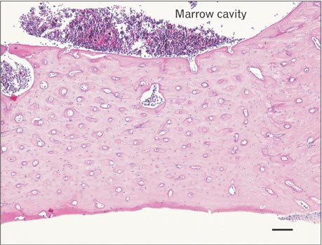
Fig. 1 Transverse section of the rat femur stained with hematoxylin and eosin. Scale bar=100 µm.
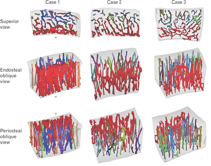
Fig. 2 Three-dimensional reconstruction of the Haversian system. Different colors represent the different networks of the Haversian and Volkmann's canals. Transparent gray shows the outline of the bone fragment. Many, large, and complex reddish networks of the Haversian and Volkmann's canals are located on the endosteal side, while relatively simple networks are located on the periosteal side. All images have the same magnification. a)Endosteal side.
Cited by 2 articles
-
Three-dimensional microstructures of the intracortical canals in the animal model of osteoporosis
Shin-Hyo Lee, Jeong-Nam Kim, Kang-Jae Shin, Ki-Seok Koh, Wu-Chul Song
Anat Cell Biol. 2020;53(2):162-168. doi: 10.5115/acb.19.189.Real-Color Volume Models Made from Real-Color Sectioned Images of Visible Korean
Beom Sun Chung, Jin Seo Park
J Korean Med Sci. 2019;34(10):. doi: 10.3346/jkms.2019.34.e86.
Reference
-
1. Kessel RG, Kardon RH. Tissues and organs: a text-atlas of scanning microscopy. New York: W.H. Freeman and Co.;1979.2. Ovalle WK, Nahirney PC. Netter's essential histology. Philadelphia: Elsevier;2008. p. 146.3. Mescher AL. Junqueira's basic histology. 13th ed. New York: McGrawHill;2013. p. 145–148.4. Tanaka M, Yamashita E, Anwar RB, Yamada K, Ohshima H, Nomura S, Ejiri S. Radiological and histologic studies of the mandibular cortex of ovariectomized monkeys. Oral Surg Oral Med Oral Pathol Oral Radiol Endod. 2011; 111:372–380.5. Particelli F, Mecozzi L, Beraudi A, Montesi M, Baruffaldi F, Viceconti M. A comparison between micro-CT and histology for the evaluation of cortical bone: effect of polymethylmethacrylate embedding on structural parameters. J Microsc. 2012; 245:302–310.6. Britz HM, Jokihaara J, Leppänen OV, Järvinen T, Cooper DM. 3D visualization and quantification of rat cortical bone porosity using a desktop micro-CT system: a case study in the tibia. J Microsc. 2010; 240:32–37.7. Cooper DM, Matyas JR, Katzenberg MA, Hallgrimsson B. Comparison of microcomputed tomographic and microradiographic measurements of cortical bone porosity. Calcif Tissue Int. 2004; 74:437–447.8. Fiala JC. Reconstruct: a free editor for serial section microscopy. J Microsc. 2005; 218(Pt 1):52–61.9. Pazzaglia UE, Zarattini G, Giacomini D, Rodella L, Menti AM, Feltrin G. Morphometric analysis of the canal system of cortical bone: An experimental study in the rabbit femur carried out with standard histology and micro-CT. Anat Histol Embryol. 2010; 39:17–26.
- Full Text Links
-
- Actions
-
Cited
- CITED
-
- Close
- Share
-
- Similar articles
-
- Three-dimensional microstructures of the intracortical canals in the animal model of osteoporosis
- Multiple Central Osteoma Of The Mandible: Report Of A Case
- Quantitative Analysis of New Bone Formation in Various Bone Grafts by In-Vivo Tetracycline Labeling
- Angiogenesis in Distraction Osteogenesis
- A Clinical Experience of Frontal Periosteal Osteoma: 20 Cases
