Primary Histiocytic Sarcoma of the Central Nervous System
- Affiliations
-
- 1Department of Internal Medicine, Asan Medical Center, University of Ulsan College of Medicine, Seoul, Korea.
- 2Department of Pathology Asan Medical Center, University of Ulsan College of Medicine, Seoul, Korea.
- 3Department of Oncology, Asan Medical Center, University of Ulsan College of Medicine, Seoul, Korea. csuh@amc.seoul.kr
- 4Departmens of Neurology, Asan Medical Center, University of Ulsan College of Medicine, Seoul, Korea.
- 5Department of Radiology and Research Institute of Radiology, Asan Medical Center, University of Ulsan College of Medicine, Seoul, Korea.
- KMID: 2132812
- DOI: http://doi.org/10.4143/crt.2013.163
Abstract
- Histiocytic sarcoma is a type of lymphoma that rarely involves the central nervous system (CNS). Its rarity can easily lead to a misdiagnosis. We describe a patient with primary CNS histocytic sarcoma involving the cerebral hemisphere and spinal cord, who had been initially misdiagnosed as demyelinating disease. Two biopsies were necessary before a correct diagnosis was made. A histologic examination showed bizarre shaped histiocytes with larger nuclei and nuclear atypia. The cells were positive for CD68, CD163, and S-100 protein. As a resection was not feasible due to multifocality, he was treated with highdose methotrexate, but showed no response. As a result, he was switched to high dose cytarabine; but again, showed no response. The patient died 2 months from the start of chemotherapy and 8 months from the onset of symptoms. Since few patients with this condition have been described and histopathology is difficult to diagnose, suspicion of the disease is essential.
MeSH Terms
Figure
-
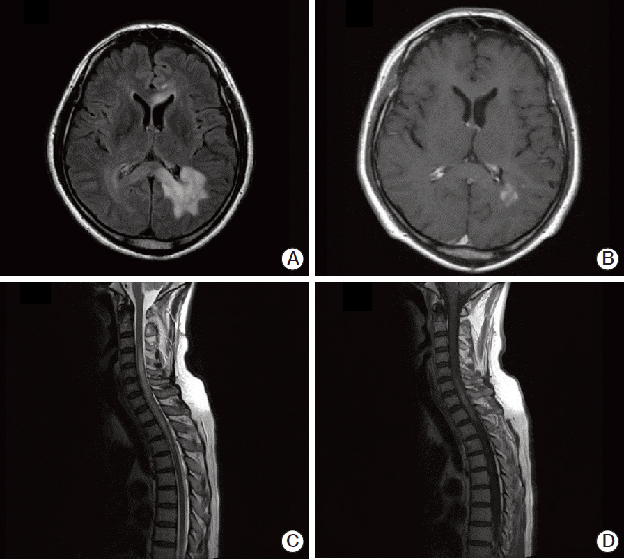
Fig. 1. Brain (A, B) and spinal (C, D) magnetic resonance imaging (MRI) of the patient. (A) Brain FLAIR MRI showing high signal intensity in the left periventricular and deep white matter of the left parietal lobe. (B) T1-weighted axial enhancement of the brain, showing subtle enhancement of the lesion. (C) Sagittal T2-weighted spinal MRI, showing diffusely increased signal intensity lesion with mild cord enlargement in the lower level of C3 through the upper level of T5. (D) Sagittal T1-weighted MRI, showing patchy enhancement of the spinal cord.
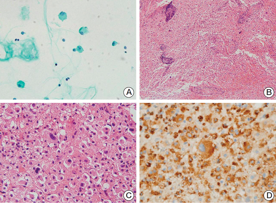
Fig. 2. Cytologic and histologic features of the first cerebrospinal fluid (CSF) aspiration and biopsy of left parieto-occipital lesion. (A) Central nervous system smear, showing a few large cells with abundant cytoplasm and large nuclei (Giemsa staining, ×400). (B) The large cells in the solid sheet from the biopsy were similar to the cells in the CSF smear. Vessels are cuffed by mature lymphocytes (H&E staining, ×40). (C) A few bizarre cells, larger than the adjacent cells, were observed (H&E staining, ×200). (D) The cells, including the bizarre cells, were positive for CD68, consistent with histiocytes (×400).

Fig. 3. Histologic features of left parieto-occipital lesion from the second biopsy. (A) Large cells were arranged in a solid sheet, with a few intermixed larger bizarre cells, similar to findings in the first biopsy specimen. The nuclei of the background cells showed slight pleomorphism and coarse chromatin with no or small nucleoli. The cytoplasm was eosinophilic and cell membranes were not well-defined (H&E staining, ×400). (B-D) The cells were positive for CD68 (B) and CD163 (C), but negative for glial fibrillary acidic protein (D), consistent with histiocytic differentiation (×200).
Reference
-
References
1. Swerdlow SH, Campo E, Harris NL, Jaffe ES, Pileri SA, Stein H, et al. WHO classification of tumours of haematopoietic and lymphoid tissues. 4th ed. Lyon: International Agency for Research on Cancer;2008.2. Gill-Samra S, Ng T, Dexter M, Wong M, Nahar N, Allsopp K, et al. Histiocytic sarcoma of the brain. J Clin Neurosci. 2012; 19:1456–8.
Article3. Toshkezi G, Edalat F, O'Hara C, Delalle I, Chin LS. Primary intramedullary histiocytic sarcoma. World Neurosurg. 2010; 74:523–7.
Article4. Bell SL, Hanzely Z, Alakandy LM, Jackson R, Stewart W. Primary meningeal histiocytic sarcoma: a report of two unusual cases. Neuropathol Appl Neurobiol. 2012; 38:111–4.
Article5. Devic P, Androdias-Condemine G, Streichenberger N, Berger F, Honnorat J, Broussolle E, et al. Histiocytic sarcoma of the central nervous system: a challenging diagnosis. QJM. 2012; 105:77–9.
Article6. Wang J, Li T, Chen H, Liu Q. A case of primary central nervous system histiocytic sarcoma. Clin Neurol Neurosurg. 2012; 114:1074–6.
Article7. Torres CF, Korones DN, Powers JM, Vadasz AG. Primary leptomeningeal histiocytic lymphoma in a young child. Med Pediatr Oncol. 1996; 27:547–50.
Article8. Cheuk W, Walford N, Lou J, Lee AK, Fung CF, Au KH, et al. Primary histiocytic lymphoma of the central nervous system: a neoplasm frequently overshadowed by a prominent inflammatory component. Am J Surg Pathol. 2001; 25:1372–9.9. Sun W, Nordberg ML, Fowler MR. Histiocytic sarcoma involving the central nervous system: clinical, immunohistochemical, and molecular genetic studies of a case with review of the literature. Am J Surg Pathol. 2003; 27:258–65.10. Cao M, Eshoa C, Schultz C, Black J, Zu Y, Chang CC. Primary central nervous system histiocytic sarcoma with relapse to mediastinum: a case report and review of the literature. Arch Pathol Lab Med. 2007; 131:301–5.
Article11. Almefty RO, Tyree TL, Fusco DJ, Coons SW, Nakaji P. Primary histiocytic sarcoma of the brain mimicking cerebral abscess. J Neurosurg Pediatr. 2013; 12:251–7.
Article12. Wu W, Tanrivermis Sayit A, Vinters HV, Pope W, Mirsadraei L, Said J. Primary central nervous system histiocytic sarcoma presenting as a postradiation sarcoma: case report and literature review. Hum Pathol. 2013; 44:1177–83.
Article13. Laviv Y, Zagzag D, Fichman-Horn S, Michowitz S. Primary central nervous system histiocytic sarcoma. Brain Tumor Pathol. 2013; 30:192–5.
Article14. Gomi K, Tanaka M, Yoshida M, Ito S, Sonoda M, Iwasaki F, et al. Primary cerebellar histiocytic sarcoma in a 17-month-old girl. J Neurosurg Pediatr. 2012; 10:126–9.
Article15. Louis DN, Ohgaki H, Wiestler OD, Cavenee WK. WHO classification of tumours of the central nervous system. 4th ed. Lyon: International Agency for Research on Cancer;2007.
- Full Text Links
-
- Actions
-
Cited
- CITED
-
- Close
- Share
-
- Similar articles
-
- A Rare Case of Isolated Central Nervous System Neoplasm With Histiocytic Features
- A Rare Case of Isolated Central Nervous System Neoplasm With Histiocytic Features
- Primary Epithelioid Hemangioendothelioma of the Central Nervous System: A Case Report
- Primary Intramedullary Spinal Sarcoma : A Case Report and Review of the Current Literatures
- Histiocytic Sarcoma of Rectum: A Case Report
