A Novel Synthetic Compound 3-Amino-3-(4-Fluoro-Phenyl)-1H-Quinoline-2,4-Dione (KR22332) Exerts a Radioprotective Effect via the Inhibition of Mitochondrial Dysfunction and Generation of Reactive Oxygen Species
- Affiliations
-
- 1Department of Otolaryngology-Head and Neck Surgery, Yonsei University Wonju College of Medicine, Wonju, Korea.
- 2Department of Otolaryngology, School of Medicine, Ajou University, Suwon, Korea. ostium@ajou.ac.kr
- 3Center for Cell Death Regulating Biodrug, School of Medicine, Ajou University, Suwon, Korea.
- 4Bio-Organic Science Division, Korea Research Institute of Chemical Technology, Daejeon, Korea.
- 5Department of Otorhinolaryngology, Yonsei Head and Neck Cancer Clinic, Yonsei University College of Medicine, Seoul, Korea.
- KMID: 2130811
- DOI: http://doi.org/10.3349/ymj.2014.55.4.886
Abstract
- PURPOSE
Acute side effects of radiation such as oral mucositis are observed in most patients. Although several potential radioprotective agents have been proposed, no effective agent has yet been identified. In this study, we investigated the effectiveness of synthetic compound 3-amino-3-(4-fluoro-phenyl)-1H-quinoline-2,4-dione (KR22332) as a radioprotective agent.
MATERIALS AND METHODS
Cell viability, apoptosis, the generation of reactive oxygen species (ROS), mitochondrial membrane potential changes, and changes in apoptosis-related signaling were examined in human keratinocyte (HaCaT).
RESULTS
KR22332 inhibited irradiation-induced apoptosis and intracellular ROS generation, and it markedly attenuated the changes in mitochondrial membrane potential in primary human keratinocytes. Moreover, KR22332 significantly reduced the protein expression levels of ataxia telangiectasia mutated protein, p53, and tumor necrosis factor (TNF)-alpha compared to significant increases observed after radiation treatment.
CONCLUSION
KR22332 significantly inhibited radiation-induced apoptosis in human keratinocytes in vitro, indicating that it might be a safe and effective treatment for the prevention of radiation-induced mucositis.
Keyword
MeSH Terms
-
Apoptosis/drug effects/physiology
Cell Line, Tumor
Cell Survival/drug effects/physiology
Humans
Keratinocytes/metabolism
Membrane Potential, Mitochondrial/drug effects/physiology
Radiation-Protective Agents/chemistry/*pharmacology
Reactive Oxygen Species/metabolism
Radiation-Protective Agents
Reactive Oxygen Species
Figure
-
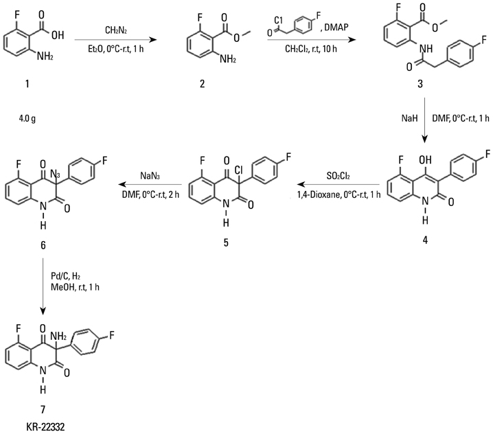
Fig. 1 Structure of 3-amino-3-(4-fluoro-phenyl)-1H-quinoline-2,4-dione (KR22332).
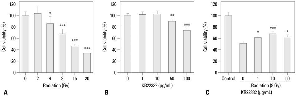
Fig. 2 Effect of KR22332 on HaCaT cell viability after radiation. HaCaT cells were exposed to various doses of radiation (0-20 Gy) or to various concentrations of KR22332 (0-100 µg/mL) with or without radiation (8 Gy). At 72 h after radiation, cell viability was measured by an MTT assay. (A) Radiation decreased cell viability in an intensity-dependent manner. (B) KR22332 alone did not show significant toxic effects on the cells until 10 µg/mL. (C) Cells were pretreated with a 8 Gy single dose of radiation followed by treatment with 1, 10, and 50 µg/mL KR22332 for 72 h. KR22332 significantly protected HaCaT cells from radiation-induced cytotoxicity in a dose-dependent manner, until 10 µg/mL. The data represent mean±SD of three independent experiments. *p<0.05, **p<0.01, ***p<0.001.
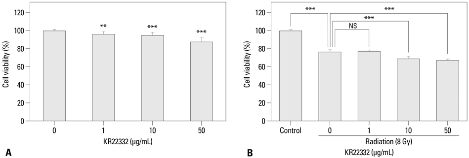
Fig. 3 Effect of KR22332 on the viability of the HNSCC cell line after treatment with radiation. (A) KR22332 did not have protective influence on the viability of HN3 cell. (B) HN3 cell line were pre-treated with 8 Gy single dose of radiation, followed by treatment with 1, 10, and 50 µg/mL KR22332 for 72 h. Then, cell viability was measured by a MTT assay. Radiation significantly decreased cell viability on HN3 cells. KR22332 treatment did not decrease anticancer effect of irradiation on the cell line. The data represent mean±SD of three independent experiments. **p<0.01, ***p<0.001. NS, not significant.
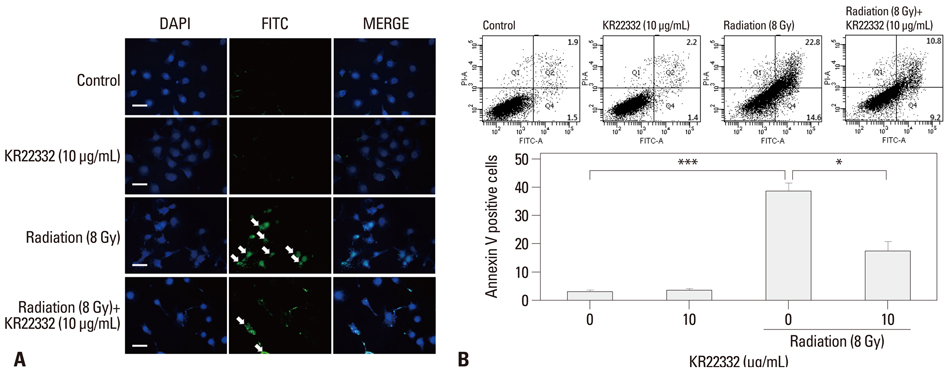
Fig. 4 Effect of KR22332 on radiation-induced apoptosis in HaCaT cells. (A) Apoptosis in HaCaT cells was determined by the TUNEL method using an in situ cell detection kit. After monolayers achieved 60-70% confluence, the cells were exposed to radiation (8 Gy), then treated with KR22332 (10 µg/mL). The cells were incubated with 50 µL of TUNEL reaction mixture (TdT and fluorescein-dUTP) and stained with Hoechst 33258 (5 µg/mL). The stained cells (arrow) were then observed under a fluorescence microscope. The TUNEL assay confirmed that radiation induced TUNEL-positive cells (arrow), while KR22332 decreased the number of TUNEL-positive cells. Scale bar=50 µm. (B) To quantify the effects of KR22332 on radiation-induced apoptosis, we used flow cytometry; annexin V-FITC and PI staining were used to analyze the percentage of apoptotic cells in radiation-treated cells (8 Gy) in the absence or presence of KR22332 (10 µg/mL) (upper). The percentage of apoptosis in each fraction is expressed as a graph (lower). The data represent mean±SD of three independent experiments. *p<0.05, ***p<0.001. DAPI, 4',6-diamidino-2-phenylindole; TUNEL, terminal deoxynucleotidyl transferase-mediated dUTP-biotin nick end labeling; V-FITC, V-fluorescein isothiocynate; PI, propidium iodide.
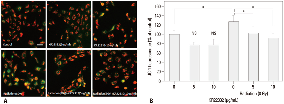
Fig. 5 Effect of KR22332 on the MMP in irradiated HaCaT cells. (A) Cells were treated with KR22332 after irradiation (8 Gy) or not, stained with JC-1, and visualized under a fluorescence microscope. KR22332 alone did not affect the MMP of cells. KR22332 stabilized the MMP in radiation-treated cells. (B) The change in the MMP was measured objectively using FACScan. The data represent mean±SD of three independent experiments. Scale bar=50 µm. *p<0.05. NS, not significant; MMP, mitochondrial membrane potential; FACS, fluorescence-activated cell sorting.
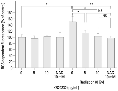
Fig. 6 Effect of KR22332 on radiation-induced ROS generation in HaCaT cells. Cells were treated with radiation (8 Gy) or KR22332 (5 µg/mL) for 72 h. The level of intracellular ROS was then measured by flow cytometry using the peroxide-sensitive fluorescent probe DCFDA. The results were calculated as a percent of the control group (not exposed to radiation). Radiation significantly increased the generation of intracellular ROS. KR22332 clearly inhibited radiation-induced intracellular ROS generation. To compare the inhibitory effect of a known inhibitor, 10 mM NAC was used to inhibit ROS generation. The data represent mean±SD of three independent experiments. *p<0.05; **p<0.01. NS, not significant; ROS, reactive oxygen species; NAC, N-acetyl-L-cysteine; DCFDA, 5-(and 6)-carboxyl-20,70-dichlorodihydro fluorescein diacetate.
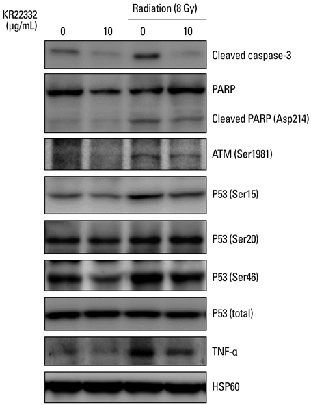
Fig. 7 Effect of KR22332 on radiation-induced apoptosis and the phosphorylation of ATM-p53 and TNF-α in HaCaT cells. The cells were incubated for 72 h after treatment with/without radiation and KR22332. Cell lysates were collected, electrophoresed through an SDS-polyacrylamide gel, and subjected to Western blot analysis with antibodies against cleaved caspase-3, cleaved PARP, ATM, p53, and TNF-α. KR22332 reduced the phosphorylation of ATM, p53 (Ser 15 and 46), and TNF-α that had been augmented by irradiation in HaCaT cells. TNF, tumor necrosis factor; PARP, poly (ADP-ribose) polymerase; ATM, ataxia telangiectasia mutated; HSP, heat shock protein.
Reference
-
1. Sonis ST, Fey EG. Oral complications of cancer therapy. Oncology (Williston Park). 2002; 16:680–686.2. Denham JW, Hauer-Jensen M. The radiotherapeutic injury--a complex "wound". Radiother Oncol. 2002; 63:129–145.3. Rose-Ped AM, Bellm LA, Epstein JB, Trotti A, Gwede C, Fuchs HJ. Complications of radiation therapy for head and neck cancers. The patient's perspective. Cancer Nurs. 2002; 25:461–467.4. Dörr W, Hamilton CS, Boyd T, Reed B, Denham JW. Radiation-induced changes in cellularity and proliferation in human oral mucosa. Int J Radiat Oncol Biol Phys. 2002; 52:911–917.
Article5. Shin YS, Song SJ, Kang SU, Hwang HS, Choi JW, Lee BH, et al. A novel synthetic compound, 3-amino-3-(4-fluoro-phenyl)-1H-quinoline-2,4-dione, inhibits cisplatin-induced hearing loss by the suppression of reactive oxygen species: in vitro and in vivo study. Neuroscience. 2012; 232C:1–12.
Article6. Chang JW, Park KH, Hwang HS, Shin YS, Oh YT, Kim CH. Protective effects of Korean red ginseng against radiation-induced apoptosis in human HaCaT keratinocytes. J Radiat Res. 2014; 55:245–256.
Article7. Spielberger R, Stiff P, Bensinger W, Gentile T, Weisdorf D, Kewalramani T, et al. Palifermin for oral mucositis after intensive therapy for hematologic cancers. N Engl J Med. 2004; 351:2590–2598.
Article8. Buentzel J, Micke O, Adamietz IA, Monnier A, Glatzel M, de Vries A. Intravenous amifostine during chemoradiotherapy for head-and-neck cancer: a randomized placebo-controlled phase III study. Int J Radiat Oncol Biol Phys. 2006; 64:684–691.
Article9. Donetti E, Bedoni M, Capone P, Gualerzi A, Tartaglia G, Sforza C. An in vitro model of human oral explants to study early effects of radiation mucositis. Eur J Oral Sci. 2009; 117:169–174.
Article10. Connolly L, Lasarev M, Jordan R, Schwartz JL, Turker MS. Atm haploinsufficiency does not affect ionizing radiation mutagenesis in solid mouse tissues. Radiat Res. 2006; 166(1 Pt 1):39–46.
Article11. Tobita T, Izumi K, Feinberg SE. Development of an in vitro model for radiation-induced effects on oral keratinocytes. Int J Oral Maxillofac Surg. 2010; 39:364–370.
Article12. Naidu MU, Ramana GV, Rani PU, Mohan IK, Suman A, Roy P. Chemotherapy-induced and/or radiation therapy-induced oral mucositis--complicating the treatment of cancer. Neoplasia. 2004; 6:423–431.
Article13. Chung YM, Park SH, Tsai WB, Wang SY, Ikeda MA, Berek JS, et al. FOXO3 signalling links ATM to the p53 apoptotic pathway following DNA damage. Nat Commun. 2012; 3:1000.
Article14. Korwek Z, Sewastianik T, Bielak-Zmijewska A, Mosieniak G, Alster O, Moreno-Villanueva M, et al. Inhibition of ATM blocks the etoposide-induced DNA damage response and apoptosis of resting human T cells. DNA Repair (Amst). 2012; 11:864–873.
Article15. Kang SU, Lee BS, Lee SH, Baek SJ, Shin YS, Kim CH. Expression of NSAID-activated gene-1 by EGCG in head and neck cancer: involvement of ATM-dependent p53 expression. J Nutr Biochem. 2013; 24:986–999.
Article16. Makovski A, Yaffe E, Shpungin S, Nir U. Down-regulation of Fer induces ROS levels accompanied by ATM and p53 activation in colon carcinoma cells. Cell Signal. 2012; 24:1369–1374.
Article17. Inagaki-Ohara K, Yada S, Takamura N, Reaves M, Yu X, Liu E, et al. p53-dependent radiation-induced crypt intestinal epithelial cells apoptosis is mediated in part through TNF-TNFR1 system. Oncogene. 2001; 20:812–818.18. Kim JJ, Lee SB, Park JK, Yoo YD. TNF-alpha-induced ROS production triggering apoptosis is directly linked to Romo1 and Bcl-X(L). Cell Death Differ. 2010; 17:1420–1434.19. Curra M, Martins MA, Lauxen IS, Pellicioli AC, Sant'Ana Filho M, Pavesi VC, et al. Effect of topical chamomile on immunohistochemical levels of IL-1β and TNF-α in 5-fluorouracil-induced oral mucositis in hamsters. Cancer Chemother Pharmacol. 2013; 71:293–299.
Article20. Gao MC, Jia XD, Wu QF, Cheng Y, Chen FR, Zhang J. Silencing Prx1 and/or Prx5 sensitizes human esophageal cancer cells to ionizing radiation and increases apoptosis via intracellular ROS accumulation. Acta Pharmacol Sin. 2011; 32:528–536.
Article
- Full Text Links
-
- Actions
-
Cited
- CITED
-
- Close
- Share
-
- Similar articles
-
- Epilepsy, Reactive Oxygen Species and Mitochondria
- Inhibition of Oxidative Tissue Damage and Mitochondrial Dysfunction by Glycyrrhizin in the 1-Methyl-4-phenyl-1,2,3,6-tetrahydropyridine Mouse Model of Parkinson's Disease
- Differential Inhibition of MPP+- or 6-Hydroxydopamine-induced Cell Viability Loss in PC12 Cells by Trifluoperazine and W-7
- Cytotoxic Mechanism of FK506 on Human T Lymphocytes
- The role of mitochondrial DNA mutation on neurodegenerative diseases
