Orthodontic treatment of a transposed maxillary canine and first premolar in a young patient with Class III malocclusion
- Affiliations
-
- 1Department of Neuroscience, University of Padova, Padova, Italy. antonio.gracco@unipd.it
- 2Post Graduate at School of Orthodontics, University of Ferrara, Ferrara, Italy.
- 3Department of Orthodontics, University of Padova, Padova, Italy.
- KMID: 2130594
- DOI: http://doi.org/10.4041/kjod.2015.45.6.322
Abstract
- A 12-year-old girl was referred to our clinic for evaluation of an unaesthetic dental appearance. All permanent teeth were erupted, while the deciduous maxillary right canine was retained. Cone-beam computed tomography revealed a complete transposition of the maxillary left canine and first premolar involving both the crowns and the roots. Initial cephalometric analysis showed a skeletal Class III pattern, with a slight maxillary retrusion and a compensated proclination of the upper incisors. The patient's teeth were considered to be in the correct position; therefore, we decided to attempt treatment by correcting the transposition and using only orthodontic compensation of the skeletal Class III malocclusion. After 25 months of active orthodontic treatment, the patient had a Class I molar and canine relationship on both sides, with ideal overbite and overjet values. Her profile was improved, her lips were competent, and cephalometric evaluation showed acceptable maxillary and mandibular incisor inclinations. The final panoramic radiograph showed that good root parallelism was achieved. Two-year follow-up intraoral photography showed stable results.
MeSH Terms
Figure
-
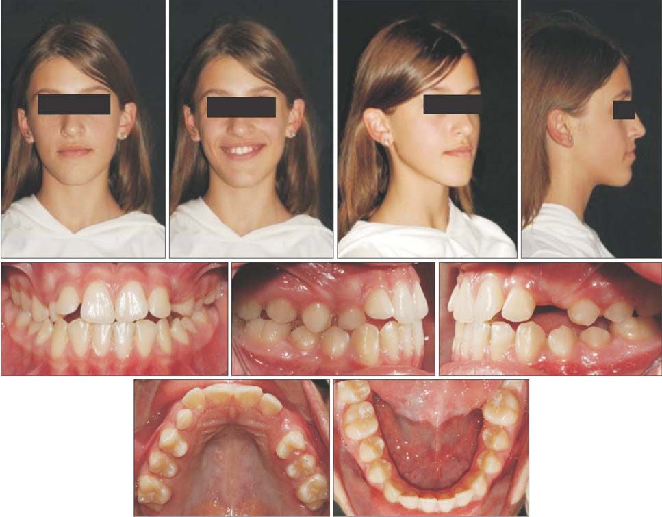
Figure 1 Pretreatment records: facial and intraoral photographs.
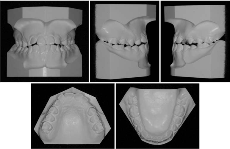
Figure 2 Pretreatment records: dental casts.
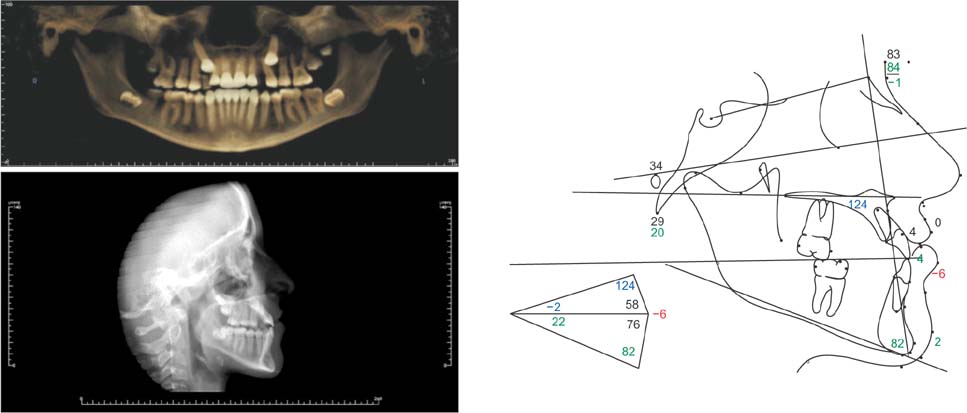
Figure 3 Pretreatment records: radiographs and cephalometric tracings.
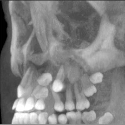
Figure 4 Three-dimensional reconstruction of the maxilla, with transposition of the left canine and first premolar.
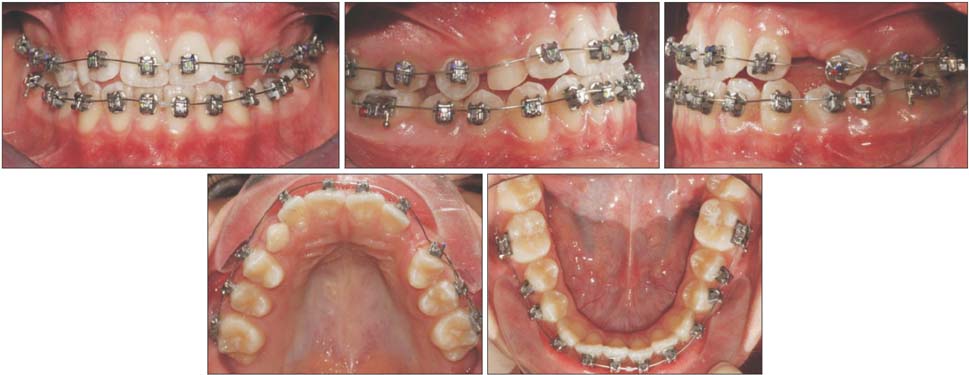
Figure 5 Intraoral photographs at bonding stage. A high-torque central incisor bracket (+17o torque value) was bonded to the first left premolar to facilitate the correction of the transposition.
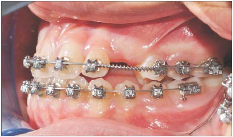
Figure 6 Intraoral photographs of the left side, showing the positive crown torque of the first premolar. The palatal cuspid of the tooth is extruded compared with the buccal cuspid of the same tooth.

Figure 7 Surgical procedure. A, Intraoral photo of the surgical exposure of the left canine; B, mucosal flap closed over the canine.
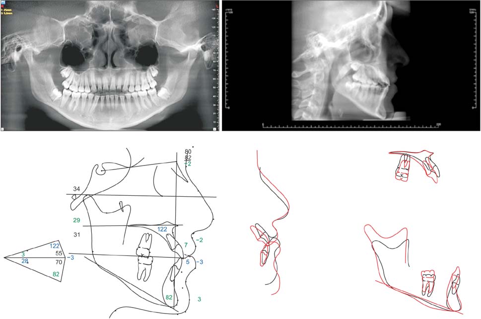
Figure 8 Post-treatment records: radiographs and cephalometric tracings (black, before treatment; red, after treatment).
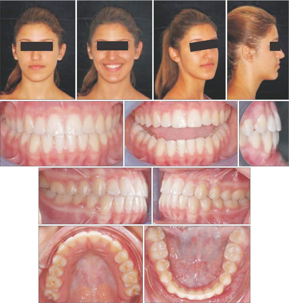
Figure 9 Post-treatment records: facial and intraoral photographs.
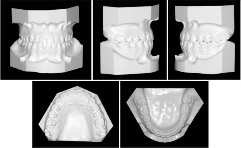
Figure 10 Post-treatment records: dental casts.
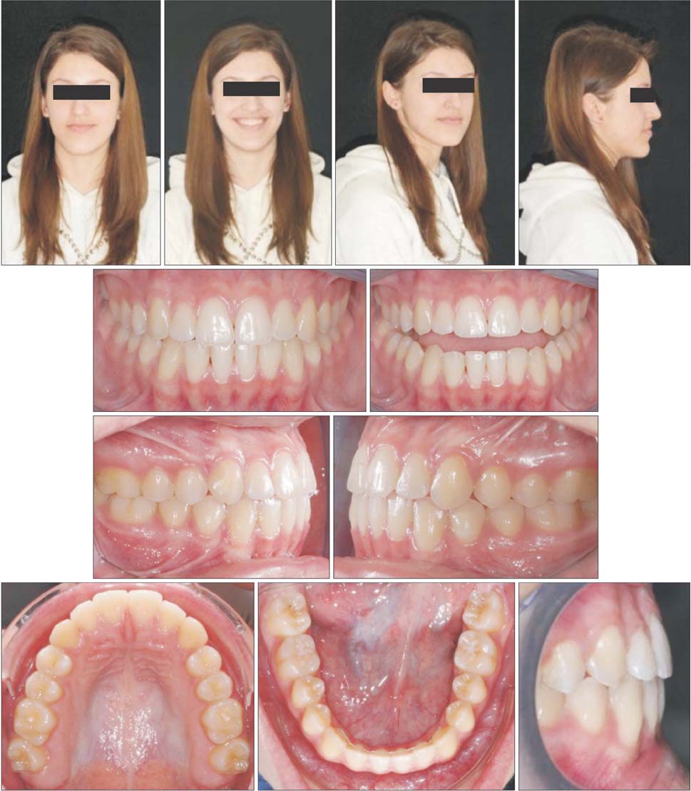
Figure 11 Two-year follow-up photographs: facial and intraoral photographs.
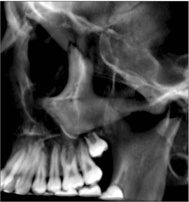
Figure 12 Three-dimensional image of the absence of progression of left first premolar (2.4) root resorption.
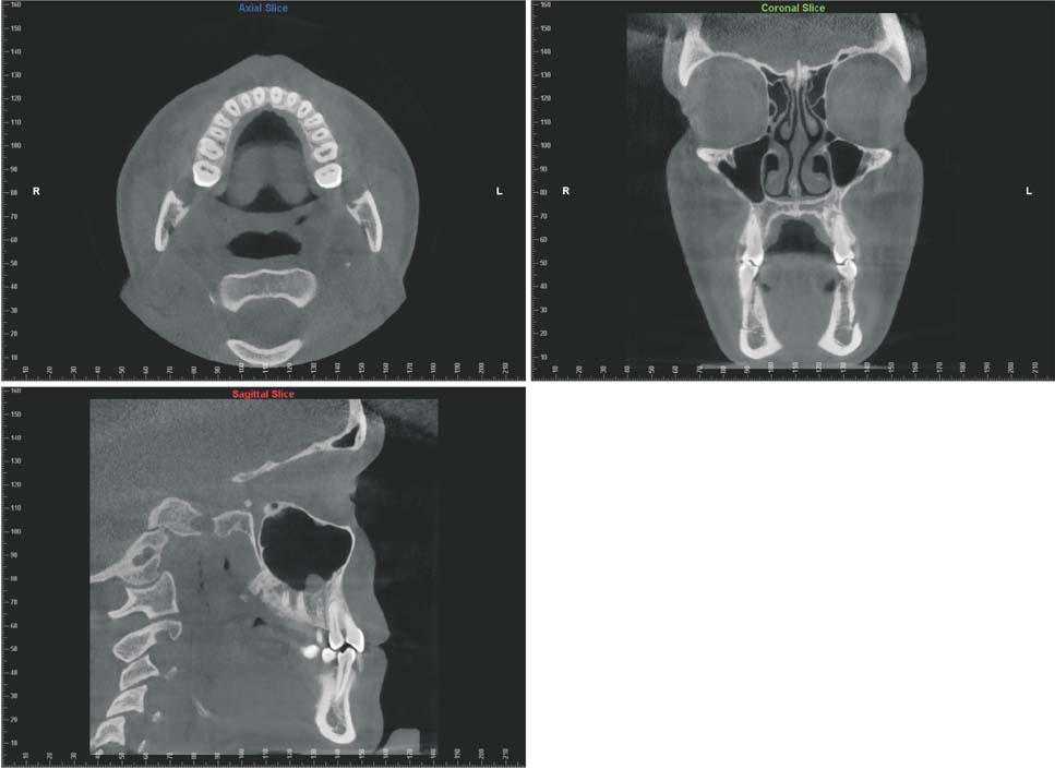
Figure 13 Three-dimensional image of root placement in maxillary medullar bone.
Cited by 1 articles
-
Long-term survival of retained deciduous mandibular second molars and maxillary canine incorporated into final occlusion
Soonshin Hwang, Yoon Jeong Choi, Chooryung J. Chung, Kyung-Ho Kim
Korean J Orthod. 2017;47(5):323-333. doi: 10.4041/kjod.2017.47.5.323.
Reference
-
1. Babacan H, Kiliç B, Biçakçi A. Maxillary canine-first premolar transposition in the permanent dentition. Angle Orthod. 2008; 78:954–960.
Article2. Sato K, Yokozeki M, Takagi T, Moriyama K. An orthodontic case of transposition of the upper right canine and first premolar. Angle Orthod. 2002; 72:275–278.3. Cho SY, Chu V, Ki Y. A retrospective study on 69 cases of maxillary tooth transposition. J Oral Sci. 2012; 54:197–203.
Article4. Chattopadhyay A, Srinivas K. Transposition of teeth and genetic etiology. Angle Orthod. 1996; 66:147–152.5. Peck L, Peck S, Attia Y. Maxillary canine-first premolar transposition, associated dental anomalies and genetic basis. Angle Orthod. 1993; 63:99–109.6. Feichtinger C, Rossiwall B, Wunderer H. Canine transposition as autosomal recessive trait in an inbred kindred. J Dent Res. 1977; 56:1449–1452.
Article7. Svinhufvud E, Myllärniemi S, Norio R. Dominant inheritance of tooth malpositions and their association to hypodontia. Clin Genet. 1988; 34:373–381.
Article8. Laptook T, Silling G. Canine transposition: approaches to treatment. J Am Dent Assoc. 1983; 107:746–748.9. Farret MM, Farret MM, Farret AM, Hollweg H. Unusual orthodontic approach to a maxillary caninepremolar transposition and a missing lateral incisor with long-term follow-up. Am J Orthod Dentofacial Orthop. 2012; 142:690–697.
Article10. Ely NJ, Sherriff M, Cobourne MT. Dental transposition as a disorder of genetic origin. Eur J Orthod. 2006; 28:145–151.
Article11. Ciarlantini R, Melsen B. Maxillary tooth transposition: correct or accept. Am J Orthod Dentofacial Orthop. 2007; 132:385–394.
Article12. Giacomet F, Araújo MT. Orthodontic correction of a maxillary canine-first premolar transposition. Am J Orthod Dentofacial Orthop. 2009; 136:115–123.
Article13. Nishimura K, Nakao K, Aoki T, Fuyamada M, Saito K, Goto S. Orthodontic correction of a transposed maxillary canine and first premolar in the permanent dentition. Am J Orthod Dentofacial Orthop. 2012; 142:524–533.
Article14. Weeks EC, Power SM. The presentations and management of transposed teeth. Br Dent J. 1996; 181:421–424.
Article15. Filho LC, Cardoso MA, An TL, Bertoz FA. Maxillary canine-first premolar transposition. Restoring normal tooth order with segmented mechanics. Angle Orthod. 2007; 77:167–175.16. Nakajima A, Sameshima GT, Arai Y, Homme Y, Shimizu N, Dougherty H Sr. Two- and three-dimensional orthodontic imaging using limited cone beam-computed tomography. Angle Orthod. 2005; 75:895–903.17. Walker L, Enciso R, Mah J. Three-dimensional localization of maxillary canines with cone-beam computed tomography. Am J Orthod Dentofacial Orthop. 2005; 128:418–423.
Article18. Maverna R, Gracco A. Different diagnostic tools for the localization of impacted maxillary canines: clinical considerations. Prog Orthod. 2007; 8:28–44.
- Full Text Links
-
- Actions
-
Cited
- CITED
-
- Close
- Share
-
- Similar articles
-
- Effect of maxillary premolar extraction on transverse arch dimension in Class III surgical-orthodontic treatment
- A study on maxillary basal bone morphology in skeletal Class III malocclusion requiring orthognathic surgery
- Study of horizontal skeletal pattern and dental arch in skeletal Class III malocclusion patients
- Correction of a maxillary canine-first premolar transposition using mini-implant anchorage
- Treatment of Class II malocclusions with upper second molar extraction
