J Adv Prosthodont.
2015 Apr;7(2):146-150. 10.4047/jap.2015.7.2.146.
The effect of zirconia framework design on the failure of all-ceramic crown under static loading
- Affiliations
-
- 1Prosthodontics Department, Faculty of Dentistry, Mahidol University, Bangkok, Thailand. s.urapepon@gmail.com
- 2Takua-thung Hospital, Phang-nga, Thailand.
- KMID: 2118245
- DOI: http://doi.org/10.4047/jap.2015.7.2.146
Abstract
- PURPOSE
This in vitro study aimed to compare the failure load and failure characteristics of two different zirconia framework designs of premolar crowns when subjected to static loading.
MATERIALS AND METHODS
Two types of zirconia frameworks, conventional 0.5 mm even thickness framework design (EV) and 0.8 mm cutback of full contour crown anatomy design (CB), were made for 10 samples each. The veneer porcelain was added on under polycarbonate shell crown made by vacuum of full contour crown to obtain the same total thickness of the experiment crowns. The crowns were cemented onto the Cobalt-Chromium die. The dies were tilted 45 degrees from the vertical plane to obtain the shear force to the cusp when loading. All crowns were loaded at the lingual incline of the buccal cusp until fracture using a universal testing machine with cross-head speed 0.5 mm/min. The load to fracture values (N) was recorded and statistically analyzed by independent sample t-test.
RESULTS
The mean and standard deviations of the failure load were 1,170.1 +/- 90.9 N for EV design and 1,450.4 +/- 175.7 N for CB design. A significant difference in the compressive failure load was found (P<.05). For the failure characteristic, the EV design was found only cohesive failures within veneering porcelain, while the CB design found more failures through the zirconia framework (8 from 10 samples).
CONCLUSION
There was a significant difference in the failure load between two designs, and the design of the framework influences failure characteristic of zirconia crown.
Figure
-

Fig. 1 Framework and veneering thickness of the experimental crowns in this study. (A) even thickness design, (B) cutback design.
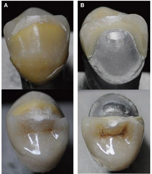
Fig. 2 Failure pattern of the experimental crowns. (A) Cohesive failure within veneering porcelain, (B) Fracture through the framework.
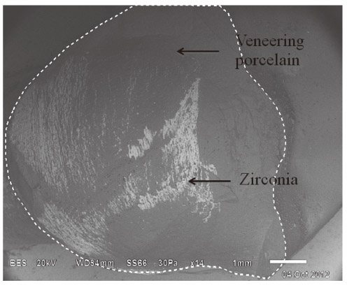
Fig. 3 Scanning electron micrograph of the fracture surface: dot line is representing all fracture area, the gray surface is veneering porcelain layer and white surface is the exposed zirconia framework.
Reference
-
1. Raigrodski AJ, Chiche GJ, Potiket N, Hochstedler JL, Mohamed SE, Billiot S, Mercante DE. The efficacy of posterior three-unit zirconium-oxide-based ceramic fixed partial dental prostheses: a prospective clinical pilot study. J Prosthet Dent. 2006; 96:237–244.2. Rosentritt M, Steiger D, Behr M, Handel G, Kolbeck C. Influence of substructure design and spacer settings on the in vitro performance of molar zirconia crowns. J Dent. 2009; 37:978–983.3. Coelho PG, Silva NR, Bonfante EA, Guess PC, Rekow ED, Thompson VP. Fatigue testing of two porcelain-zirconia all-ceramic crown systems. Dent Mater. 2009; 25:1122–1127.4. Marchack BW, Futatsuki Y, Marchack CB, White SN. Customization of milled zirconia copings for all-ceramic crowns: a clinical report. J Prosthet Dent. 2008; 99:169–173.5. Shoher I, Whiteman AE. Reinforced porcelain system: a new concept in ceramometal restorations. J Prosthet Dent. 1983; 50:489–496.6. Marker JC, Goodkind RJ, Gerberich WW. The compressive strength of nonprecious versus precious ceramometal restorations with various frame designs. J Prosthet Dent. 1986; 55:560–567.7. Sailer I, Fehér A, Filser F, Lüthy H, Gauckler LJ, Schärer P, Franz Hämmerle CH. Prospective clinical study of zirconia posterior fixed partial dentures: 3-year follow-up. Quintessence Int. 2006; 37:685–693.8. Al-Amleh B1, Lyons K, Swain M. Clinical trials in zirconia: a systematic review. J Oral Rehabil. 2010; 37:641–652.9. Tinschert J, Schulze KA, Natt G, Latzke P, Heussen N, Spiekermann H. Clinical behavior of zirconia-based fixed partial dentures made of DC-Zirkon: 3-year results. Int J Prosthodont. 2008; 21:217–222.10. Larsson C1, El Madhoun S, Wennerberg A, Vult von Steyern P. Fracture strength of yttria-stabilized tetragonal zirconia polycrystals crowns with different design: an in vitro study. Clin Oral Implants Res. 2012; 23:820–826.11. Swain MV. Unstable cracking (chipping) of veneering porcelain on all-ceramic dental crowns and fixed partial dentures. Acta Biomater. 2009; 5:1668–1677.12. Richerson D, Richerson DW, Lee WE. Modern ceramic engineering: properties, processing and use in design. 3rd ed. Boca Raton: Taylor and Francis Group;2006.13. Bonfante EA, da Silva NR, Coelho PG, Bayardo-González DE, Thompson VP, Bonfante G. Effect of framework design on crown failure. Eur J Oral Sci. 2009; 117:194–199.14. Khers SC, Carpenter CW, Vetter JD, Staley RN. Anatomy of cusps of posterior teeth and their fracture potential. J Prosthet Dent. 1990; 64:139–147.15. Pinho ST, D'avila CG, Camanho PP, Iannucci L, Robinson P. Failure models and criteria for FRP under in-plane or threedimensional stress states including shear non-linearity. NASA/TM-2005-213530, L-19089.16. Cavel WT, Kelsey WP, Blankenau RJ. An in vivo study of cuspal fracture. J Prosthet Dent. 1985; 53:38–42.17. Farah JW, Craig RG. Distribution of stresses in porcelainfused-to-metal and porcelain jacket crowns. J Dent Res. 1975; 54:255–261.18. Craig RG, el-Ebrashi MK, Peyton FA. Stress distribution in porcelain-fused-to-gold crowns and preparations constructed with photoelastic plastics. J Dent Res. 1971; 50:1278–1283.19. Nally JN, Farah JW, Craig RG. Experimental stress analysis of dental restorations. IX. Two-dimensional photoelastic stress analysis of porcelain bonded to gold crowns. J Prosthet Dent. 1971; 25:307–316.20. Lawn BR, Pajares A, Zhang Y, Deng Y, Polack MA, Lloyd IK, Rekow ED, Thompson VP. Materials design in the performance of all-ceramic crowns. Biomaterials. 2004; 25:2885–2892.21. Kleinfelder JW, Ludwigt K. Maximal bite force in patients with reduced periodontal tissue support with and without splinting. J Periodontol. 2002; 73:1184–1187.
- Full Text Links
-
- Actions
-
Cited
- CITED
-
- Close
- Share
-
- Similar articles
-
- Fracture strength of zirconia monolithic crowns and metal-ceramic crowns after cyclic loading and thermocycling
- The effect of different cooling rates and coping thicknesses on the failure load of zirconia-ceramic crowns after fatigue loading
- Fracture strength of zirconia monolithic crowns
- Fracture strength of zirconia ceramic crowns according to tooth position
- An alternative method to convert fractured metal ceramic surveyed crown into a complete contour zirconia surveyed crown using CAD-CAM technology under anticancer treatments: a clinical report
