Chonnam Med J.
2010 Dec;46(3):195-198. 10.4068/cmj.2010.46.3.195.
Surgical Correction of Lid Retraction with a Silicone Sponge in Congenital Fibrosis of the Extraocular Muscles
- Affiliations
-
- 1Department of Ophthalmology, Chonnam National University Medical School & Hospital, Gwangju, Korea. exo70@naver.com
- KMID: 2116776
- DOI: http://doi.org/10.4068/cmj.2010.46.3.195
Abstract
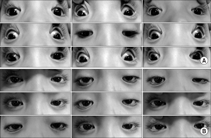
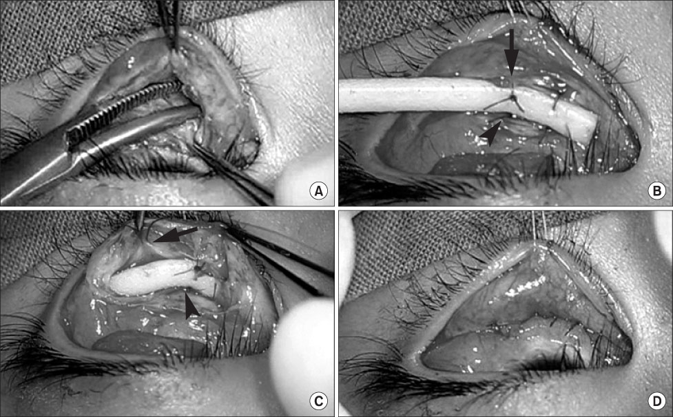
- A 5-year-old boy who was diagnosed with congenital fibrosis of the extraocular muscles (CFEOM) presented with bilateral lower eyelid retraction after strabismus surgery. To correct the lower eyelid retraction, a silicone sponge was used as a spacer; it was sutured to the inferior border of the tarsus and the lower eyelid retractor. At the 3-month postoperative visit, both lower eyelid margins were at the inferior corneal limbus. No complications or recurrence was noted at 1 year after surgery. The silicone sponge served as a good spacer for correction of lower lid retraction after strabismus surgery in a child with CFEOM.
Keyword
MeSH Terms
Figure
Reference
-
1. Doxanas MT, Dryden RM. The use of sclera in the treatment of dysthyroid eyelid retraction. Ophthalmology. 1981. 88:887–894.
Article2. Baylis HI, Perman KI, Fett DR, Sutcliffe RT. Autogenous auricular cartilage grafting for lower eyelid retraction. Ophthal Plast Reconstr Surg. 1985. 1:23–27.
Article3. Kersten RC, Kulwin DR, Levartovsky S, Tiradellis H, Tse DT. Management of lower-lid retraction with hard-palate mucosa grafting. Arch Ophthalmol. 1990. 108:1339–1343.
Article4. Downes RN, Jordan K. The surgical management of dysthyroid related eyelid retraction using Mersilene mesh. Eye (Lond). 1989. 3:385–390.
Article5. Tan J, Olver J, Wright M, Maini R, Neoh C, Dickinson AJ. The use of porous polyethylene (Medpor) lower eyelid spacers in lid heightening and stabilisation. Br J Ophthalmol. 2004. 88:1197–1200.
Article6. Lincoff HA, Baras I, Mclean J. Modifications to the custodies procedure for retinal detachment. Arch Ophthalmol. 1965. 73:160–163.7. Apt L, Axelrod RN. Generalized fibrosis of the extraocular muscles. Am J Ophthalmol. 1978. 85:822–829.
Article8. Engle EC. The genetic basis of complex strabismus. Pediatr Res. 2006. 59:343–348.
Article9. Engle EC, Goumnerov BC, McKeown CA, Schatz M, Johns DR, Porter JD, et al. Oculomotor nerve and muscle abnormalities in congenital fibrosis of the extraocular muscles. Ann Neurol. 1997. 41:314–325.
Article10. Lee YJ, Khwarg SI. Polytetrafluoroethylene as a spacer graft for the correction of lower eyelid retraction. Korean J Ophthalmol. 2005. 19:247–251.
Article
- Full Text Links
- Actions
-
Cited
- CITED
-
- Close
- Share
- Similar articles
-
- A Case Report of the Congenital Fibrosis of Extraocular Muscles
- Surgical Correction of Lower Lid Retraction Using The Scleral Spacer
- Two Cases of Congenital Fibrosis of the Extraocular Muleles
- Congenital Generalized Fibrosis of Extraocular Muscles in a Family
- Lower Eyelid Epiblepharon Associated with Lower Eyelid Retraction


