Brain Tumor Res Treat.
2015 Oct;3(2):89-94. 10.14791/btrt.2015.3.2.89.
Analysis of Circulating Endostatin and Vascular Endothelial Growth Factor in Patients with Pituitary Adenoma Treated by Stereotactic Radiosurgery: A Preliminary Study
- Affiliations
-
- 1Department of Neurosurgery, Kyungpook National University Hospital, Daegu, Korea. nsdoctor@naver.com
- KMID: 2114653
- DOI: http://doi.org/10.14791/btrt.2015.3.2.89
Abstract
- BACKGROUND
The purpose of this study was to investigate plasma levels of endostatin and vascular endothelial growth factor (VEGF) in normal subjects and in patients with pituitary adenoma and to evaluate change in these levels following stereotactic radiosurgery (SRS) for pituitary adenoma.
METHODS
Peripheral venous blood was collected from five patients with pituitary adenoma before SRS using Gamma Knife and at the 1 week and 1 month follow-up visits. Plasma endostatin and VEGF levels were measured using commercially available enzyme-linked immunosorbent assay kits. Peripheral blood samples were obtained from 10 healthy volunteers as controls.
RESULTS
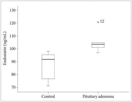
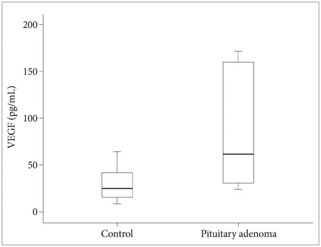
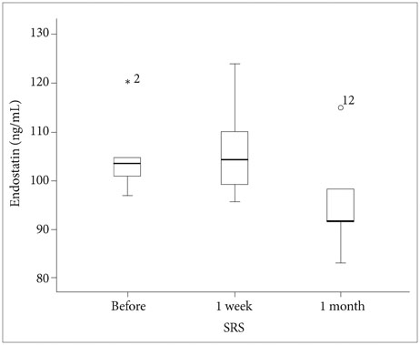
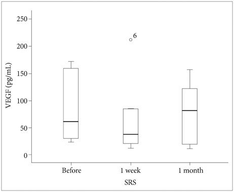
Mean baseline plasma endostatin level (105.3 ng/mL, range, 97.0-120.2 ng/mL) in patients with pituitary adenoma was higher than that of the healthy controls (86.6 ng/mL, range, 71.3-98.2 ng/mL) (p=0.001). Mean plasma VEGF level was 89.5 pg/mL (range, 24.1-171.8 pg/mL) in patients with pituitary adenoma at baseline and 29.3 pg/mL (range, 9.2-64.3 pg/mL) in the control group (p=0.050). Plasma endostatin level changed to 106.6 ng/mL 1 week after SRS and decreased to 95.9 ng/mL after 1 month. Plasma VEGF level following SRS decreased to 74.1 pg/mL after 1 week and 79.0 pg/mL after 1 month. There was a trend toward decreased plasma endostatin and VEGF concentrations 1 month after SRS compared to baseline levels (p=0.195, p=0.812, respectively).
CONCLUSION
Plasma endostatin and VEGF levels in patients with pituitary adenoma were significantly elevated over controls at baseline, which decreased from baseline to 1 month after SRS for pituitary adenomas.
Keyword
MeSH Terms
Figure
Reference
-
1. Sánchez-Ortiga R, Sánchez-Tejada L, Moreno-Perez O, Riesgo P, Niveiro M, Picó Alfonso AM. Over-expression of vascular endothelial growth factor in pituitary adenomas is associated with extrasellar growth and recurrence. Pituitary. 2013; 16:370–377.
Article2. Sheehan JP, Starke RM, Mathieu D, et al. Gamma Knife radiosurgery for the management of nonfunctioning pituitary adenomas: a multicenter study. J Neurosurg. 2013; 119:446–456.
Article3. Gruszka A, Kunert-Radek J, Pawlikowski M, Stepien H. Serum endostatin levels are elevated and correlate with serum vascular endothelial growth factor levels in patients with pituitary adenomas. Pituitary. 2005; 8:163–168.
Article4. Komorowski J, Jankewicz J, Stepień H. Vascular endothelial growth factor (VEGF), basic fibroblast growth factor (bFGF) and soluble interleukin-2 receptor (sIL-2R) concentrations in peripheral blood as markers of pituitary tumours. Cytobios. 2000; 101:151–159.5. Turner HE, Harris AL, Melmed S, Wass JA. Angiogenesis in endocrine tumors. Endocr Rev. 2003; 24:600–632.
Article6. O'Reilly MS, Boehm T, Shing Y, et al. Endostatin: an endogenous inhibitor of angiogenesis and tumor growth. Cell. 1997; 88:277–285.7. Lee CC, Kano H, Yang HC, et al. Initial Gamma Knife radiosurgery for nonfunctioning pituitary adenomas. J Neurosurg. 2014; 120:647–654.
Article8. Kraft A, Weindel K, Ochs A, et al. Vascular endothelial growth factor in the sera and effusions of patients with malignant and nonmalignant disease. Cancer. 1999; 85:178–187.
Article9. Vermeulen S, Young R, Li F, et al. A comparison of single fraction radiosurgery tumor control and toxicity in the treatment of basal and nonbasal meningiomas. Stereotact Funct Neurosurg. 1999; 72:Suppl 1. 60–66.
Article10. Nonoguchi N, Miyatake S, Fukumoto M, et al. The distribution of vascular endothelial growth factor-producing cells in clinical radiation necrosis of the brain: pathological consideration of their potential roles. J Neurooncol. 2011; 105:423–431.
Article11. Feldman AL, Alexander HR Jr, Bartlett DL, et al. A prospective analysis of plasma endostatin levels in colorectal cancer patients with liver metastases. Ann Surg Oncol. 2001; 8:741–745.
Article12. Feldman AL, Tamarkin L, Paciotti GF, et al. Serum endostatin levels are elevated and correlate with serum vascular endothelial growth factor levels in patients with stage IV clear cell renal cancer. Clin Cancer Res. 2000; 6:4628–4634.13. Zucker S, Mirza H, Conner CE, et al. Vascular endothelial growth factor induces tissue factor and matrix metalloproteinase production in endothelial cells: conversion of prothrombin to thrombin results in progelatinase A activation and cell proliferation. Int J Cancer. 1998; 75:780–786.
Article14. Páez Pereda M, Ledda MF, Goldberg V, et al. High levels of matrix metalloproteinases regulate proliferation and hormone secretion in pituitary cells. J Clin Endocrinol Metab. 2000; 85:263–269.
Article15. Stepien HM, Kołomecki K, Pasieka Z, Komorowski J, Stepień T, Kuzdak K. Angiogenesis of endocrine gland tumours--new molecular targets in diagnostics and therapy. Eur J Endocrinol. 2002; 146:143–151.
Article16. O'Sullivan EP, Woods C, Glynn N, et al. The natural history of surgically treated but radiotherapy-naïve nonfunctioning pituitary adenomas. Clin Endocrinol (Oxf). 2009; 71:709–714.
- Full Text Links
- Actions
-
Cited
- CITED
-
- Close
- Share
- Similar articles
-
- Influence of vascular endothelial growth factor (VEGF) and endostatin on coronary artery lesions in Kawasaki disease
- Usefulness of circulating vascular endothelial growth factor and neutrophil elastase as diagnostic markers of disseminated intravascular coagulation in non-cancer patients
- Change in Plasma Vascular Endothelial Growth Factor after Gamma Knife Radiosurgery for Meningioma: A Preliminary Study
- The Roles of Vascular Endothelial Growth Factor, Angiostatin, and Endostatin in Nasal Polyp Development
- Pituitary Epithelioid Osteosarcoma after Gamma-knife Surgery of a Pituitary Adenoma

