J Korean Orthop Assoc.
2015 Feb;50(1):37-44. 10.4055/jkoa.2015.50.1.37.
Comparison of Histological and Biochemical Findings with Magnetic Resonance Imaging on the Degeneration Severity of Meniscus
- Affiliations
-
- 1Department of Orthopedic Surgery, International St. Mary's Hospital, Catholic Kwandong University College of Medicine, Incheon, Korea. drchaeos@gmail.com
- KMID: 2106736
- DOI: http://doi.org/10.4055/jkoa.2015.50.1.37
Abstract
- PURPOSE
The aim of this study is to determine whether degeneration severities of meniscus assessed using magnetic resonance imaging (MRI) would well estimate those assessed using histological and biochemical examinations.
MATERIALS AND METHODS
Seven lateral menisci from knees with osteoarthritis undergoing total knee arthroplasty (study group) and five from normal controls (control group) were examined for this study. Degeneration severities of the menisci were graded using MRI, histologic and biochemical examinations of the menisci were then performed. Comparative analyses of MRI grading and results of histological/biochemical examinations of the menisci were performed in each group. In addition, comparative analyses of histological/biochemical conditions were performed between specimens of the study group and the control group showing grade 0 on MRI.
RESULTS
All specimens from the control group showed grade 0 on MRI and their histology was also grade 0. In addition, no significant differences in biochemical results were observed among the specimens of the control group. In the lateral meniscus from the study group showing degeneration on MRI it was found that the water and proteoglycan contents increased with increasing grade of degeneration whereas the collagen content decreased. The meniscus specimens of the control group and the study group showing grade 0 on MRI had similar histologic findings but had different biochemical properties. The grade I, II degenerations on MRI were not well matched with the histologic findings in the study group.
CONCLUSION
Severities of meniscus degeneration on MRI did not well reflect the histologic findings of the meniscus. This finding may be due to the water content of the meniscus. The factors of the high signal intensity of the degenerated lateral meniscus on MRI may be due to the decreased component of collagen and increased proteoglycan. Our findings suggested that caution should be taken when the severities of meniscus degeneration on MRI are attributed to histologic severities of degenerated meniscus.
Keyword
MeSH Terms
Figure
-

Figure 1 Scout magnetic resonance imaging of the degenerated lateral meniscus. 1, 0-10 degree; 6, 51-60 degree; 12, 111-120 degree; 18, 171-180 degree of the lateral meniscus.
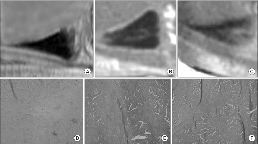
Figure 2 Lotysch's grading of the degenerated meniscus on magnetic resonance imaging of the experimental group. (A) Grade 0; (B) Grade I; (C) Grade II. Histology of the degeneratd lateral meniscus. (D) Stage 0 (H&E, ×100); (E) Stage 1 (H&E, ×40); (F) Stage 2 (H&E, ×40).
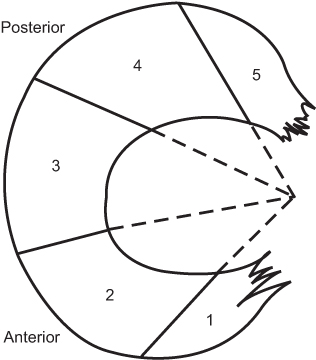
Figure 3 The preparation of the specimen that was divided into 5 regions.
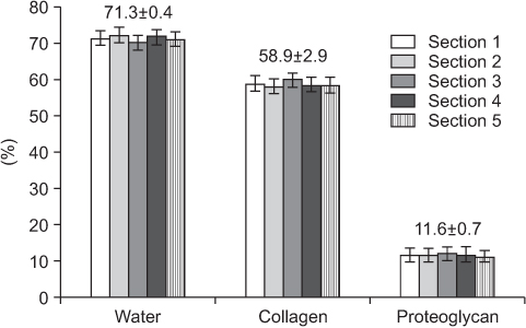
Figure 4 Biochemical analysis of the meniscus of control group according to the region.
Reference
-
1. Herwig J, Egner E, Buddecke E. Chemical changes of human knee joint menisci in various stages of degeneration. Ann Rheum Dis. 1984; 43:635–640.
Article2. McDevitt CA, Webber RJ. The ultrastructure and biochemistry of meniscal cartilage. Clin Orthop Relat Res. 1990; 252:8–18.
Article3. Stoller DW, Martin C, Crues JV 3rd, Kaplan L, Mink JH. Meniscal tears: pathologic correlation with MR imaging. Radiology. 1987; 163:731–735.
Article4. Hajek PC, Gylys-Morin VM, Baker LL, Sartoris DJ, Haghighi P, Resnick D. The high signal intensity meniscus of the knee. Magnetic resonance evaluation and in vivo correlation. Invest Radiol. 1987; 22:883–890.
Article5. Hauger O, Frank LR, Boutin RD, et al. Characterization of the "red zone" of knee meniscus: MR imaging and histologic correlation. Radiology. 2000; 217:193–200.
Article6. Hodler J, Haghighi P, Pathria MN, Trudell D, Resnick D. Meniscal changes in the elderly: correlation of MR imaging and histologic findings. Radiology. 1992; 184:221–225.
Article7. Quinn SF, Muus C, Sara A, Estrada J, Walling A. Meniscal tears: pathologic correlation with MR imaging. Radiology. 1988; 166:580–581.
Article8. Lotysch M, Mink J, Crues JV, Schwartz SA. Magnetic resonance imaging in the detection of meniscal injuries. Magn Reson Imaging. 1986; 4:185.
Article9. De Smet AA, Norris MA, Yandow DR, Graf BK, Keene JS. Diagnosis of meniscal tears of the knee with MR imaging: effect of observer variation and sample size on sensitivity and specificity. AJR Am J Roentgenol. 1993; 160:555–559.
Article10. Herman LJ, Beltran J. Pitfalls in MR imaging of the knee. Radiology. 1988; 167:775–781.
Article11. Reicher MA, Hartzman S, Duckwiler GR, Bassett LW, Anderson LJ, Gold RH. Meniscal injuries: detection using MR imaging. Radiology. 1986; 159:753–757.
Article12. Chan PS, Kneeland JB, Gannon FH, Luchetti WT, Herzog RJ. Identification of the vascular and avascular zones of the human meniscus using magnetic resonance imaging: correlation with histology. Arthroscopy. 1998; 14:820–823.
Article13. Raunest J, Oberle K, Loehnert J, Hoetzinger H. The clinical value of magnetic resonance imaging in the evaluation of meniscal disorders. J Bone Joint Surg Am. 1991; 73:11–16.
Article14. Negendank WG, Fernandez-Madrid FR, Heilbrun LK, Teitge RA. Magnetic resonance imaging of meniscal degeneration in asymptomatic knees. J Orthop Res. 1990; 8:311–320.
Article15. Hough AJ Jr, Webber RJ. Pathology of the meniscus. Clin Orthop Relat Res. 1990; 252:32–40.
Article16. Ferrer-Roca O, Vilalta C. Lesions of the meniscus. Part I: macroscopic and histologic findings. Clin Orthop Relat Res. 1980; 146:289–300.
