Bilateral Hypertrophic Olivary Degeneration in Wilson Disease
- Affiliations
-
- 1Department of Neuroradiology, Leipzig University Hospital, Leipzig 04103, Germany. josephin.otto@medizin.uni-leipzig.de
- 2Department of Neurology, Leipzig University Hospital, Leipzig 04103, Germany.
- KMID: 1482793
- DOI: http://doi.org/10.3348/kjr.2013.14.2.316
Abstract
- Hypertrophic olivary degeneration resulting from lesions of the dento-rubro-olivary pathway, also called Guillain-Mollaret-triangle, has been described previously in a number of cases. Reports about bilateral hypertrophic olivary degeneration of the inferior olivary nuclei are very limited, and the magnetic resonance imaging findings of hypertrophic olivary degeneration in Wilson disease have not yet been described to the best of our knowledge. Herein, we present the first report of bilateral hypertrophic olivary degeneration diagnosed by magnetic resonance imaging in a patient suffering from Wilson disease.
MeSH Terms
Figure
-
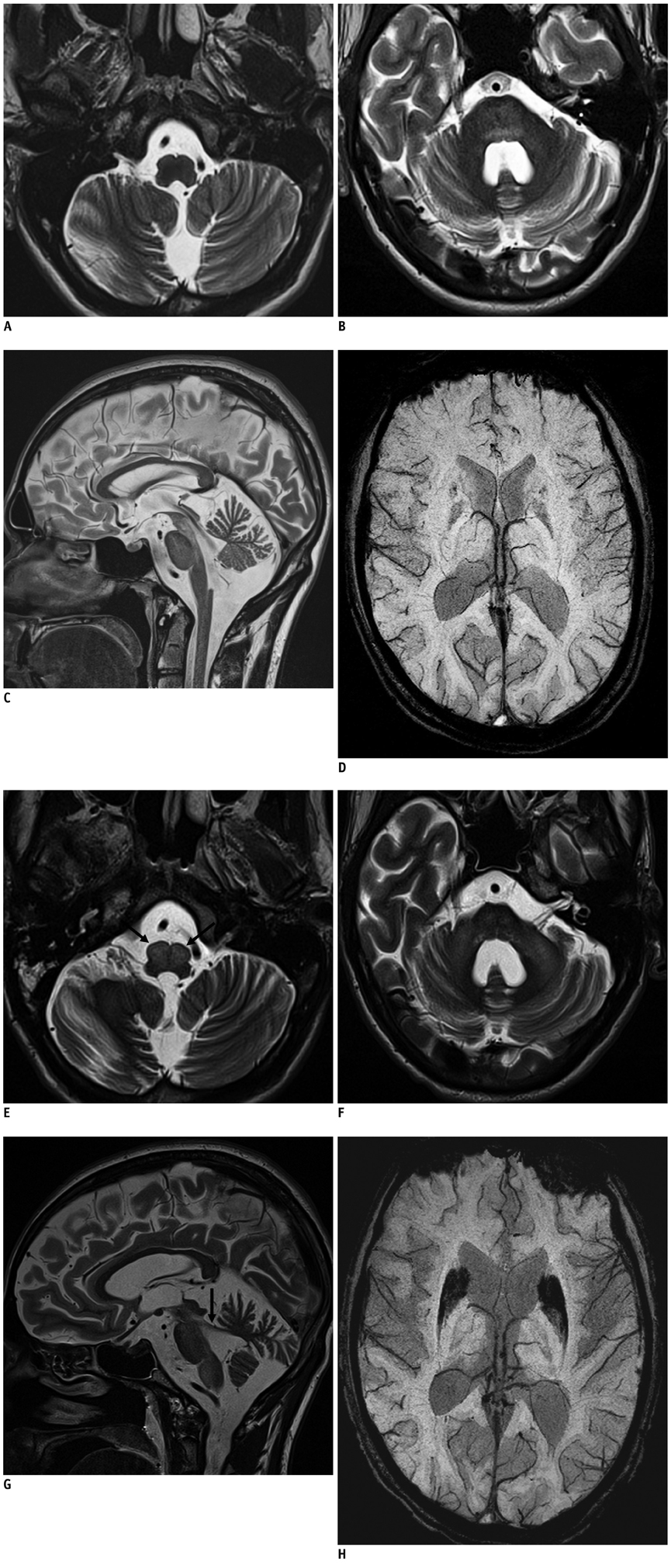
Fig. 1 Bilateral hypertrophic olivary degeneration in Wilson disease (WD). 23-year-old man suffering from WD with initial MRI findings. A. Transverse T2WI (TR/TE: 6000/96 ms, FA = 120°) at level of medulla oblongata without signs of hypertrophy or increased signal intensity of inferior olivary nuclei on both sides. B. Transverse T2WI at level of pons: symmetrical hyperintensity of pontine tegmentum. C. Midsagittal T2WI: hyperintense midbrain with atrophy of cerebellum. D. Transverse SWI (TR/TE: 28/20 ms, FA = 15°) at level of basal ganglia shows low signal intensity of heads of caudate nuclei and putamina. SWI = susceptibility weighted image, T2WI = T2-weighted image MRI findings of same patient one year later. E. Transverse T2WI (TR/TE: 6000/96 ms, FA = 120°) at level of medulla oblongata: bilateral hyperintense and hypertrophic inferior olivary nuclei (arrows). F. Transverse T2WI at level of pons: symmetrical hyperintensity and atrophy of medial cerebellar peduncles and pons. G. Parasagittal T2WI: hyperintense midbrain, pons, medulla; atrophy of superior cerebellar peduncle (arrow) and cerebellum. H. Transverse SWI (TR/TE: 28/20 ms, FA = 15°) at level of basal ganglia: considerable volume and ongoing signal loss in heads of caudate nuclei and putamina. SWI = susceptibility weighted image, T2WI = T2-weighted image
Cited by 1 articles
-
Holmes' Tremor Associated with Bilateral Hypertrophic Olivary Degeneration Following Brain Stem Hemorrhage: A Case Report
Min Kyu Kim, Byung Moon Cho, Se-Hyuck Park, Dae Young Yoon
J Cerebrovasc Endovasc Neurosurg. 2014;16(3):299-302. doi: 10.7461/jcen.2014.16.3.299.
Reference
-
1. Hornyak M, Osborn AG, Couldwell WT. Hypertrophic olivary degeneration after surgical removal of cavernous malformations of the brain stem: report of four cases and review of the literature. Acta Neurochir (Wien). 2008. 150:149–156. discussion 156.2. Kitajima M, Korogi Y, Shimomura O, Sakamoto Y, Hirai T, Miyayama H, et al. Hypertrophic olivary degeneration: MR imaging and pathologic findings. Radiology. 1994. 192:539–543.3. Asal N, Yılmaz O, Turan A, Yiğit H, Duymuş M, Tekin E. Hypertrophic olivary degeneration after pontine hemorrhage. Neuroradiology. 2012. 54:413–415.4. Goyal M, Versnick E, Tuite P, Cyr JS, Kucharczyk W, Montanera W, et al. Hypertrophic olivary degeneration: metaanalysis of the temporal evolution of MR findings. AJNR Am J Neuroradiol. 2000. 21:1073–1077.5. Phatouros CC, McConachie NS. Hypertrophic olivary degeneration: case report in a child. Pediatr Radiol. 1998. 28:830–831.6. Goto N, Kaneko M. Olivary enlargement: chronological and morphometric analyses. Acta Neuropathol. 1981. 54:275–282.7. Salamon-Murayama N, Russell EJ, Rabin BM. Diagnosis please. Case 17: hypertrophic olivary degeneration secondary to pontine hemorrhage. Radiology. 1999. 213:814–817.8. Birbamer G, Buchberger W, Felber S, Aichner F. MR appearance of hypertrophic olivary degeneration: temporal relationships. AJNR Am J Neuroradiol. 1992. 13:1501–1503.9. Dinçer A, Özyurt O, Kaya D, Koşak E, Öztürk C, Erzen C, et al. Diffusion tensor imaging of Guillain-Mollaret triangle in patients with hypertrophic olivary degeneration. J Neuroimaging. 2011. 21:145–151.10. Gerace C, Fele MR, Luna R, Piazza G. Neurological picture. Bilateral hypertrophic olivary degeneration. J Neurol Neurosurg Psychiatry. 2006. 77:73.11. Auffray-Calvier E, Desal HA, Naudou-Giron E, Severin-Fontana S, Cavenaile-Dolez H, Stefan A, et al. [Hypertrophic olivary degeneration. MR imaging findings and temporal evolution]. J Neuroradiol. 2005. 32:67–72.12. Tsui EY, Cheung YK, Mok CK, Yuen MK, Chan JH. Hypertrophic olivary degeneration following surgical excision of brainstem cavernous hemangioma: a case report. Clin Imaging. 1999. 23:215–217.13. van Wassenaer-van Hall HN, van den Heuvel AG, Jansen GH, Hoogenraad TU, Mali WP. Cranial MR in Wilson disease: abnormal white matter in extrapyramidal and pyramidal tracts. AJNR Am J Neuroradiol. 1995. 16:2021–2027.14. Magalhaes AC, Caramelli P, Menezes JR, Lo LS, Bacheschi LA, Barbosa ER, et al. Wilson's disease: MRI with clinical correlation. Neuroradiology. 1994. 36:97–100.15. Sinha S, Taly AB, Ravishankar S, Prashanth LK, Vasudev MK. Central pontine signal changes in Wilson's disease: distinct MRI morphology and sequential changes with de-coppering therapy. J Neuroimaging. 2007. 17:286–291.16. King AD, Walshe JM, Kendall BE, Chinn RJ, Paley MN, Wilkinson ID, et al. Cranial MR imaging in Wilson's disease. AJR Am J Roentgenol. 1996. 167:1579–1584.
- Full Text Links
-
- Actions
-
Cited
- CITED
-
- Close
- Share
-
- Similar articles
-
- Bilateral Hypertrophic Degeneration of the Inferior Olivary Nucleus secondary to Infarction of the Brainstem and Cerebellum: A Case Report
- MR Imaging of Hypertrophic Olivary Degeneration, Emphasising on its Causes and Patterns
- Bilateral Hypertrophic Olivary Degeneration with Oculopalatal Tremor after Brainstem Hemorrhage: A case report
- Bilateral Wallerian Degeneration of the Middle Cerebellar Peduncle and Unilateral Hypertrophic Olivary Degeneration Secondary to Pontine Hemorrhage: A Case Report
- Hemiplegia and Palatal Myoclonus after Hypertrophic Olivary Degeneration: A case report
