J Korean Soc Magn Reson Med.
2014 Jun;18(2):167-170. 10.13104/jksmrm.2014.18.2.167.
Medial Longitudinal Fasciculus on MRI in a Patient with Internuclear Ophthalmoparesis: A Case Report
- Affiliations
-
- 1Department of Radiology, Catholic University of Daegu School of Medicine, Daegu, Korea.
- 2Department of Radiology, Kyungpook National University Hospital, Daegu, Korea. knuhrad@yahoo.co.kr
- KMID: 2099893
- DOI: http://doi.org/10.13104/jksmrm.2014.18.2.167
Abstract
- The medial longitudinal fasciculus (MLF) is myelinated composite tract, lying near the midline, ventral to periaqueductal grey matter that plays a key role in coordinating eye movements. A lesion of the MLF results in an ipsilateral adduction deficit and a contralateral abducting nystagmus, referred to as an internuclear ophthalmoparesis. The blended tract with adjacent white matter in pons and midbrain is indistinguishable on brain imaging such as CT and MRI. Until now, to the best of our knowledge, MLF is not delineated on in vivo MRI. We present a case showing the whole connecting courses of MLF lesion on MRI in a patient with inflammatory demyelinating disorder.
Keyword
MeSH Terms
Figure
-
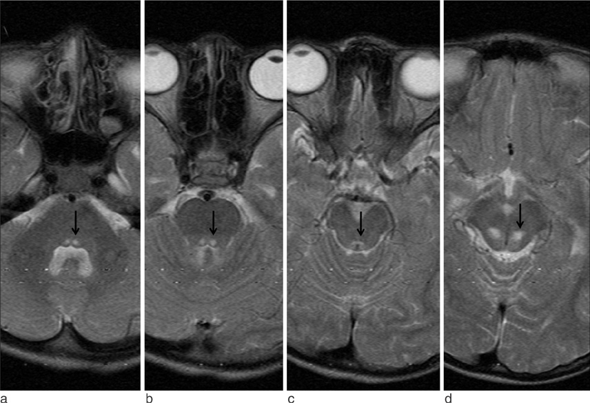
Fig. 1 T2 weighed image of MR imaging showed bilateral contiguous high signal intensities at the posterior aspect of pons and midbrain. High signal intensities of the pons represent ascending fibers from the abducent nucleus (a, arrow) to the occulomotor nucleus (d, arrow) via medial longitudinal fasciculus (b and c). The bilateral fasciculi run medially and decussated at the lower level of midbrain (c, arrows).
Reference
-
1. Standring S. Gray's anatomy. 39th ed. London: Churchill Livingstone;2004.2. Pola J, Robinson DA. An explanation of eye movements seen in internuclear ophthalmoplegia. Arch Neurol. 1976; 33:447–452.3. Highstein SM, Baker R. Excitatory termination of abducens internuclear neurons on medial rectus motoneurons: relationship to syndrome of internuclear ophthalmoplegia. J Neurophysiol. 1978; 41:1647–1661.4. Carpenter MB. Core text of Neuroanatomy. 4th Ed. Baltimore: Williams & Wilkins;1991.5. Hirose G, Furui K, Yoshioka A, Sakai K. Unilateral conjugate gaze palsy due to a lesion of the abducens nucleus. Clinical and neuroradiological correlations. J Clin Neuroophthalmol. 1993; 13:54–58.6. Frohman EM, Zhang H, Kramer PD, et al. MRI characteristics of the MLF in MS patients with chronic internuclear ophthalmoparesis. Neurology. 2001; 57:762–768.7. Keane JR. Internuclear ophthalmoplegia: unusual causes in 114 of 410 patients. Arch Neurol. 2005; 62:714–717.8. Atlas SW, Grossman RI, Savino PJ, et al. Internuclear ophthalmoplegia: MR-anatomic correlation. AJNR Am J Neuroradiol. 1987; 8:243–247.9. Napadow V, Dhond R, Kennedy D, Hui KK, Makris N. Automated brainstem co-registration (ABC) for MRI. Neuroimage. 2006; 32:1113–1119.10. Sakaie K, Takahashi M, Dimitrov I, et al. Diffusion tensor imaging the medial longitudinal fasciculus in INO: opportunities and challenges. Ann N Y Acad Sci. 2011; 1233:307–312.
- Full Text Links
-
- Actions
-
Cited
- CITED
-
- Close
- Share
-
- Similar articles
-
- A Case of Bilateral Internuclear Ophthalmoplegia
- A Case of Bilateral Internuclear Ophthalmoplegia
- A Case of Bilateral Internuclear Ophthalmoplegia with No Brain Stem Lesion
- Bilateral Internuclear Ophthalmoplegia in Tuberculous Meningits (A Report of one case)
- Wall-Eyed Monocular Internuclear Ophthalmoplegia (WEMINO) with Contraversive Ocular Tilt Reaction
