Cardiac MRI and Transthoracic Echocardiography of Left Ventricular Myocardial Noncompaction in A Patient with Congestive Heart Failure: A Case Report
- Affiliations
-
- 1Department of Radiology, Chosun University Hospital, Korea.
- 2Department of Radiology, Soonchunhyang University Hospital Bucheon, Korea. dhk0827@schmc.ac.kr
- KMID: 2097911
- DOI: http://doi.org/10.3348/jksr.2010.63.5.419
Abstract
- We report a case of a 38-year-old male presenting with new-onset dyspnea, that was diagnosed as left ventricular noncompaction by transthoracic echocardiographic and cardiac MR. The tests revealed left ventricularsystolic dysfunction with prominent trabeculations associated with deep intertrabecular recesses and an end-diastolic noncompacted to compacted ratio of 2.5 in the whole apical wall and mid-ventricular anterolateraland inferolateral walls. Delayed gadolinium contrast-enhanced MRI revealed subepicardial mid-wall hyperenhancement of the midventricular anteroseptal and inferoseptal walls, which suggested myocardial fibrosis. We review the pathophysiology, clinical characteristics, and diagnostic approach of the left ventricular noncompaction associated with congestive heart failure.
MeSH Terms
Figure
-
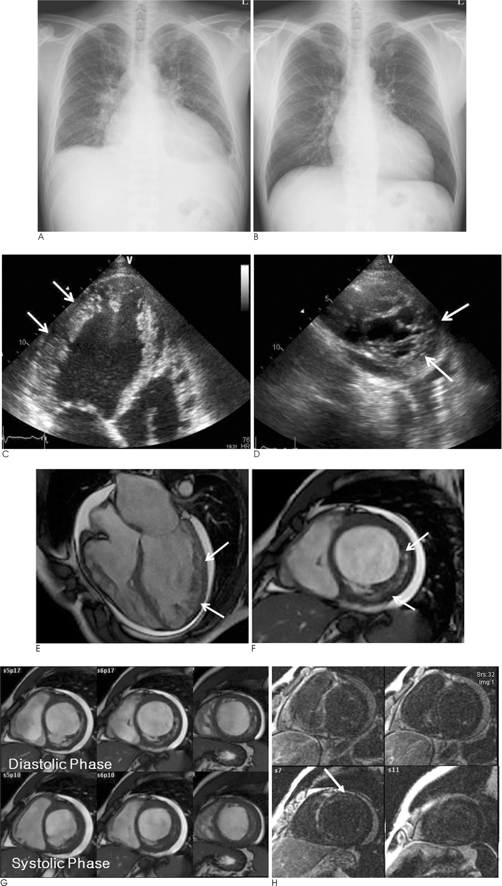
Fig. 1 A 38-year-old male with dyspnea. A. Plain chest radiograph demonstrated cardiomegaly, pulmonary congestion and mild both pleural effusion. B. Follow-up chest radiograph after adequate treatment showed improvement of pulmonary congestion and both pleural effusion. C. Apical 4-chamber view of transthoracic echocardiography showed typical deep recesses and prominent trabeculations involving the left ventricular apex and lateral wall (arrows). D. Parasternal short axis view of transthoracic echocardiography demonstrated the noncompacted myocardium of left ventricle (arrows). E. Static 4-chamber view of cine MR image showed prominent ventricular myocardial trabeculations and deep intertrabecular recesses communicating with the ventricular cavity, compromising the mid-ventricular lateral wall and the apex of the left ventricle (arrows). F. Short-axis view presented two myocardial layers with different degrees of tissue compaction (compacted and noncompacted, arrows). The end-diastolic noncompacted to compacted ratio measured 2.5. G. Short-axis cine MR images during diastolic (upper images) and systolic (lower images) phases showed global hypokinesia and prominent noncompacted myocardium. H. Basal to apical short-axis images acquired 15 minutes after the injection of the contrast agent showed poorly marginated enhancement of anterior ventricular wall.
Reference
-
1. Chin TK, Perloff JK, Williams RG, Jue K, Mohrmann R. Isolated noncompaction of left ventricular myocardium. A study of eight cases. Circulation. 1990; 82:507–513.2. Varnava AM. Isolated left ventricular non-compaction: a distinct cardiomyopathy? Heart. 2001; 86:599–600.3. Jenni R, Oechslin E, Schneider J, Attenhofer Jost C, Kaufmann PA. Echocardiographic and pathoanatomical characteristics of isolated left ventricular non-compaction: a step towards classification as a distinct cardiomyopathy. Heart. 2001; 86:666–671.4. Oechslin EN, Attenhofer Jost CH, Rojas JR, Kaufmann PA, Jenni R. Long-term follow-up of 34 adults with isolated left ventricular noncompaction: a distinct cardiomyopathy with poor prognosis. J Am Coll Cardiol. 2000; 36:493–500.5. Pignatelli RH, McMahon CJ, Dreyer WJ, Denfield SW, Price J, Belmont JW, et al. Clinical characterization of left ventricular noncompaction in children: a relatively common form of cardiomyopathy. Circulation. 2003; 108:2672–2678.6. Petersen SE, Selvanayagam JB, Wiesmann F, Robson MD, Francis JM, Anderson RH, et al. Left ventricular non-compaction: insights from cardiovascular magnetic resonance imaging. J Am Coll Cardiol. 2005; 46:101–105.7. Alhabshan F, Smallhorn JF, Golding F, Musewe N, Freedom RM, Yoo SJ. Extent of myocardial non-compaction: comparison between MRI and echocardiographic evaluation. Pediatr Radiol. 2005; 35:1147–1151.8. Richardson P, McKenna W, Bristow M, Maisch B, Mautner B, O'Connell J, et al. Report of the 1995 World Health Organization/International Society and Federation of Cardiology Task Force on the Definition and Classification of cardiomyopathies. Circulation. 1996; 93:841–842.9. Aras D, Tufekcioglu O, Ergun K, Ozeke O, Yildiz A, Topaloglu S, et al. Clinical features of isolated ventricular noncompaction in adults long-term clinical course, echocardiographic properties, and predictors of left ventricular failure. J Card Fail. 2006; 12:726–733.10. Espinola-Zavaleta N, Soto ME, Castellanos LM, Jativa-Chavez S, Keirns C. Non-compacted cardiomyopathy: clinical-echocardiographic study. Cardiovasc Ultrasound. 2006; 4:35.
- Full Text Links
-
- Actions
-
Cited
- CITED
-
- Close
- Share
-
- Similar articles
-
- A case of isolated noncompaction of the ventricular myocardium in an elderly patient
- Anesthetic experience of patient with isolated left ventricular noncompaction: a case report
- Left Ventricular Noncompaction Complicated with Myocardial Infarction with Barth Syndrome in a Newborn
- Noncompaction of Ventricular Myocardium Involving the Right Ventricle
- Isolated Right Ventricular Noncompaction Accompanied by Right Ventricular Failure
