Monolobar Caroli's Disease in Left Lobe of the Liver: A Case Report
- Affiliations
-
- 1Department of Radiology, Wonkwang University Hospital, Korea. yjyh@wonkwang.ac.kr
- KMID: 2097901
- DOI: http://doi.org/10.3348/jksr.2010.63.3.249
Abstract
- Caroli's disease is a rare congenital hepatobiliary disease characterized by multifocal segmental dilatation of the intrahepatic bile ducts and hepatic fibrosis that can cause bile duct stones, cholangitis, and cholangiocarcinoma. The disease may diffusely affect the liver or be localized to one lobe or segment. Less than 20% of all reported cases of Caroli's disease are the monolobar type. We report a case of Caroli's disease of the monolobar type, which was confined to segment 4a of the liver in a 30-year-old man. The disease was diagnosed by CT and Gd-EOB-DTPA enhanced MRI, and confirmed histopathologically after a hepatic lobectomy.
MeSH Terms
Figure
-
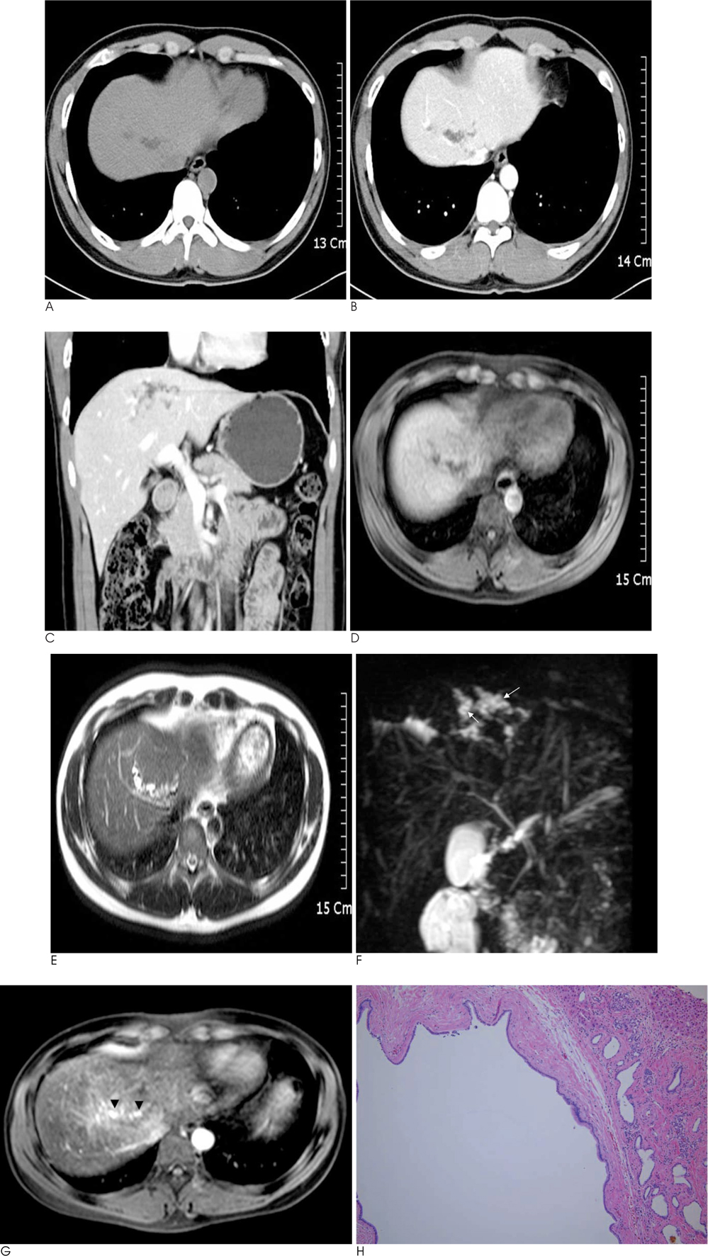
Fig. 1 A 30-year-old man with monolobar Caroli's disease in segment 4 of the liver. A-C. Non-contrast CT (A) and contrast enhanced axial (B) and coronal (C) images show focal tortuous low density tubular structures at segment 4 of the liver. No remarkable enhancing soft tissue lesion or high density stone is noted. D. T1-weighted axial image shows toutorus tubular low signal intensity lesion at segment 4a of the liver. E. T2-weighted axial image shows high signal intensity tubular structures suggestive dilated intrahepatic bile ducts and small low signal nodular foci at wall of dilated bile ducts suggestive fibrovascular bundles. F. T2-MRCP image shows lobulated high signal intensity cystic lesions with small low signal intensity nodular foci (arrows) suggesting dilated intrahepatic bile ducts and fibrovascular bundles. G. One hour delayed T1-wighted axial image after Gd-EOB-DTPA injection and 10 minutes after superparamagnetic iron oxide agent injection, shows high signal intensity contrast materials (arrowheads) in dilated intrahepatic bile ducts in segment 4 of the liver, demonstrate that the cystic lesions are dilated intrahepatic bile ducts. H. Microphotogram shows dilated lumen of intrahepatic bile ducts with surrounding inflammatory cell infiltration and mild fibrosis. There is no stone or neoplastic lesion.
Reference
-
1. Desmet VJ. Congenital diseases of intrahepatic bile ducts: variations on the theme ductal plate malformation. Hepatology. 1992; 16:1069–1083.2. Verma SK, Mitchell DG. Images: Monolobar Caroli's disease. Indian J Radiol Imaging. 2007; 17:254–256.3. Dayton MT, Longmire WP Jr, Tompkins RK. Caroli's disease: a premalignant condition? Am J Surg. 1983; 145:41–48.4. Boyle MJ, Doyle GD, McNulty JG. Monolobar Caroli's disease. Am J Gastroenterol. 1989; 84:1437–1444.5. Kassahun WT, Kahn T, Wittekind C, Moössner J, Caca K, Hauss J, et al. Caroli's disease: liver resection and liver transplantation. Experience in 33 patients. Surgery. 2005; 138:888–898.6. Kil H, Choi EY, Jeong JI, Park CS, Park SM, Kim SH, et al. A case of simple type Caroli's disease confined to right lobe of the liver. Korean J Gastroenterol. 2007; 50:271–276.7. Kim WC, Bahk YW, Kim HK. Caroli's disease. J Korean Med Assoc. 1974; 17:73–77.8. Yoo SJ, Moon YS, Lee SW, Yang JH, Park SJ, Park JW, et al. A case of simple type Caroli's disease confined to one segment of the liver. Korean J Med. 2005; 68:448–452.9. Pavone P, Laghi A, Catalano C, Materia A, Basso N, Passariello R. Caroli's disease: evaluation with MR cholangiopancreatography (MRCP). Abdom Imaging. 1996; 21:117–119.10. Bollow M, Taupitz M, Hamm B, Staks T, Wolf KJ, Weinmann HJ. Gadolinium-ethoxybenzyl-DTPA as a hepatobiliary contrast agent for use in MR cholangiography: results of an in vivo phase-I clinical evaluation. Eur Radiol. 1997; 7:126–132.
