Extrapleural Inner Thoracic Wall Lesions: Multidetector CT Findings
- Affiliations
-
- 1Department of Radiology, Soonchunhyang University Cheonan Hospital, Korea. ytokim@schca.ac.kr
- KMID: 2097897
- DOI: http://doi.org/10.3348/jksr.2010.63.3.225
Abstract
- The extrapleural space is external to the parietal pleura in the thorax. The structures within and adjacent to this region include the fat pad, endothoracic fascia, intercostal muscles, connective tissue, nerves, vessels, and ribs. Further, the space is divided into the inner and outer thoracic wall by the innermost intercostal muscle. Extrapleural lesions in the inner thoracic wall are classified as air-containing lesions, fat-containing lesions, and soft tissue-containing lesions according on their main component. Air-containing lesions include extrapleural air from direct chest trauma and extrapleural extension from pneumomediastinum. Prominent extrapleural fat is seen in decreased lung volume conditions, and can also be seen in normal individuals. Soft tissue-containing lesions include extrapleural extensions from a pleural or chest wall infection as well as tumors and extrapleural hematoma. We classify extrapleural lesions in the inner thoracic wall and illustrate their imaging findings.
MeSH Terms
Figure
-

Fig. 1 Extrapleural space in inner thoracic wall on CT. Extrapleural space is divided into inner- and outer-thoracic wall by innermost intercostal muscle.
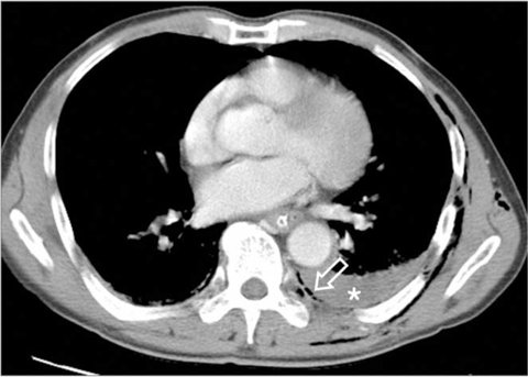
Fig. 2 Extrapleural air caused by direct chest trauma in a 75-year-old man. Axial CT scan shows extrapleural air (open arrow) external to pleural hematoma (*).

Fig. 3 Extrapleural air caused by direct chest trauma in a 55-year-old man. A. Chest radiograph shows extrapleural air and hematoma (open arrows). B. Axial CT scan with lung setting shows extrapleural hematoma and air (open arrows).
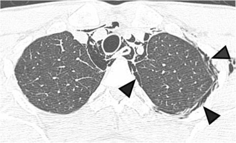
Fig. 4 Extrapleural extension from pneumomediastinum in a 29-year-old man. Axial CT scan with lung setting shows multiple air collections (arrowheads) in extrapleural space of left hemithorax and mediastinal air. Extrapleural air has internal linear opacity, so it can be differentiated from pneumothorax.
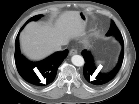
Fig. 5 Bilateral extrapleural fat in a 74-year-old man. Axial CT scan shows prominent extrapleural fat (arrows) in posterior aspect of both lower lobes.

Fig. 6 Unilateral extrapleural fat in a 63-year-old woman. Axial CT scan shows prominent extrapleural fat (*) in posterior aspect of left lower lobe.
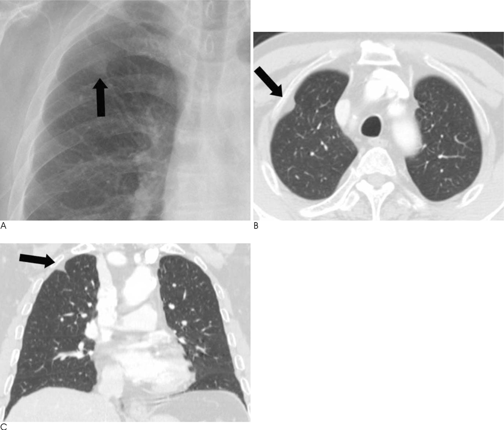
Fig. 7 Focal extrapleural fat in a 74-year-old man. A. Chest radiograph shows a focal increased opacity (arrow) with extrapleural sign in the right upper lobe. B, C. Axial (B) and coronal reformatted (C) images show focal fat deposition (arrow) in the anterolateral periphery of right upper lobe.
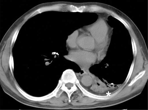
Fig. 8 Left lower lobe collapse with decreased lung volume in a 55-year-old man. Axial CT scan shows collapsed left lower lobe with traction bronchiectasis and prominent extrapleural fat (*) in the posterior aspect of left lower lobe.

Fig. 9 Lipoma at extrapleural space in a 27-year-old man. Axial scan shows a well circumscribed low attenuation mass (arrow) at the right lateral extrapleural space and invaginating the lung.
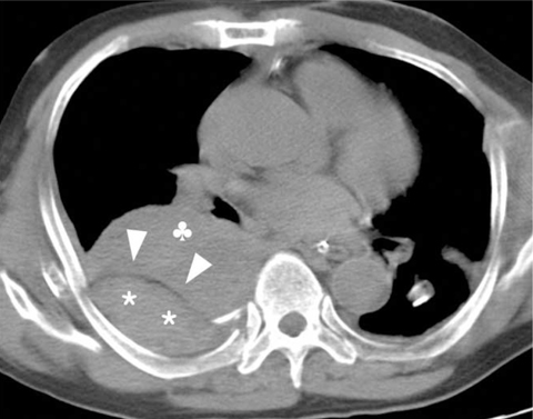
Fig. 10 Extrapleural hematoma from chest trauma in a 57-year-old man. Precontrast CT scan shows extrapleural hematoma (*) external to extrapleural fat (arrowheads), and pleural hematoma (♣) internal to extrapleural fat.

Fig. 11 Extrapleural extension from empyema in a 80-year-old man. Axial scan shows dirty increased attenuation (arrow) in extrapleural fat, posterior to the chronic empyema in right lower hemithorax. Also note prominent extrapleural fat (open arrows), pleural thickening, and calcification in left lower hemithorax.
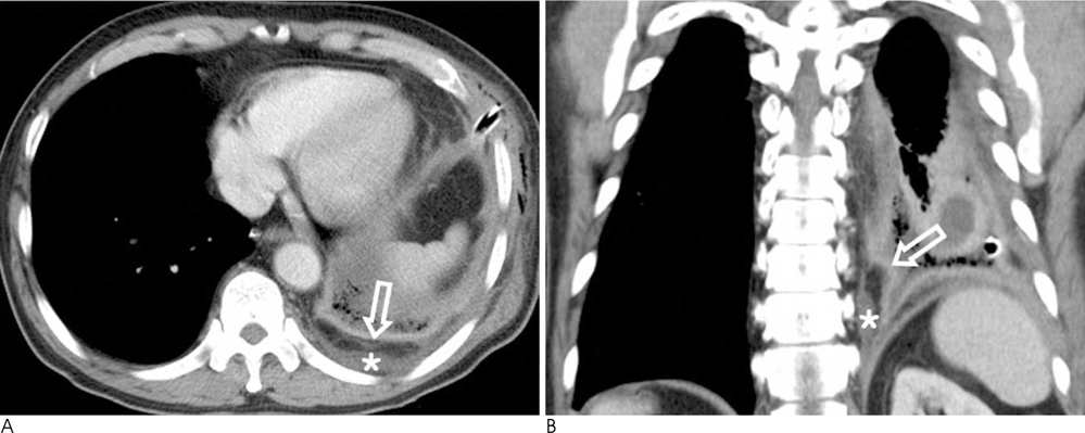
Fig. 12 Extrapleural extension from empyema in a 61-year-old man. A, B. Axial (A) and coronal reformatted (B) images show fluid collection (*) in extrapleural space external to the enhanced pleura (open arrow) and extrapleural fat.
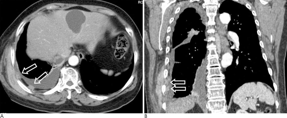
Fig. 13 Extrapleural metastasis from lung cancer in a 68-year-old woman. A, B. Axial and coronal reformatted images show enhancing nodules (open arrows) in the extrapleural space containing extrapleural fat. Also note right pleural effusion and enhancing mass in right chest wall.
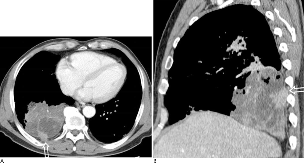
Fig. 14 Extrapleural extension from adenocarcinoma of lung in a 67-year-old man. A, B. Axial and sagittal reformatted images show dense consolidative lesion in right lower lobe. Irregular shaped enhancing mass (open arrow) in the posterior portion of consolidative lesion extends to extrapleural space in inner thoracic wall.

Fig. 15 Extrapleural metastasis from malignant pleural effusion in a 77-year-old man. A, B. Axial scans show multiple enhancing masses (open arrows) in extrapleural space containing extrapleural fat.

Fig. 16 Extrapleural and chest wall extension from malignant mesothelioma in a 64-year-old man. Axial CT scan shows enhancing nodule (open arrow) in extrapleural space containing extrapleural fat.
Reference
-
1. Im JG, Webb WR, Rosen A, Gamsu G. Costal pleura: appearances at high-resolution CT. Radiology. 1989; 171:125–131.2. Groskin SA. Selected topics in chest trauma. Radiology. 1992; 183:605–617.3. Lillard RL, Allen RP. The extrapleural air sign in pneumomediastinum. Radiology. 1965; 85:1093–1098.4. Kurihara Y, Nakajima Y, Niimi H, Arakawa H, Ishikawa T. Extrapleural air collections mimicking pneumothorax: helical CT finding. J Comput Assist Tomogr. 1997; 21:771–772.5. Fisher ER, Godwin JD. Extrapleural fat collections: pseudotumors and other confusing manifestations. AJR Am J Roentgenol. 1993; 161:47–52.6. Aquino SL, Chiles C, Oaks T. Displaced extrapleural fat as revealed by CT scanning: evidence of extrapleural hematoma. AJR Am J Roentgenol. 1997; 169:687–689.7. Waite RJ, Carbonneau RJ, Balikian JP, Umali CB, Pezzella AT, Nash G. Parietal pleural changes in empyema: appearances at CT. Radiology. 1990; 175:145–150.8. Morris BS, Maheshwari M, Chalwa A. Chest wall tuberculosis: a review of CT appearances. Br J Radiol. 2004; 77:449–457.9. Felson B. The extrapleural space. Semin Roentgenol. 1977; 12:327–333.10. Aquino SL, Chen MY, Kuo WT, Chiles C. The CT appearance of pleural and extrapleural disease in lymphoma. Clin Radiol. 1999; 54:647–650.
- Full Text Links
-
- Actions
-
Cited
- CITED
-
- Close
- Share
-
- Similar articles
-
- Traumatic Extrapleural Hematoma Mimicking Hemothorax
- Extrapleural Fat Hypertrophy in Patients with Lung Cancer: CT Findings
- Traumatic Extrapleural Hematoma Mimicking a Hemothorax
- CT Findings and Types of Tuberculous Chest Wall Abscess
- Chest Wall and Fissural Invasion of Peripheral Lung Cancer: Evaluation with HRCT
