Distalization pattern of whole maxillary dentition according to force application points
- Affiliations
-
- 1Department of Orthodontics, School of Dentistry, Yonsei University, Seoul, Korea. orthojn@yuhs.ac
- 2Division of Orthodontics, Department of Dentistry, Ewha Womans University Medical Center, Seoul, Korea.
- KMID: 2074819
- DOI: http://doi.org/10.4041/kjod.2015.45.1.20
Abstract
OBJECTIVE
The purpose of this study was to observe stress distribution and displacement patterns of the entire maxillary arch with regard to distalizing force vectors applied from interdental miniscrews.
METHODS
A standard three-dimensional finite element model was constructed to simulate the maxillary teeth, periodontal ligament, and alveolar process. The displacement of each tooth was calculated on x, y, and z axes, and the von Mises stress distribution was visualized using color-coded scales.
RESULTS
A single distalizing force at the archwire level induced lingual inclination of the anterior segment, and slight intrusive distal tipping of the posterior segment. In contrast, force at the high level of the retraction hook resulted in lingual root movement of the anterior segment, and extrusive distal translation of the posterior segment. As the force application point was located posteriorly along the archwire, the likelihood of extrusive lingual inclination of the anterior segment increased, and the vertical component of the force led to intrusion and buccal tipping of the posterior segment. Rotation of the occlusal plane was dependent on the relationship between the line of force and the possible center of resistance of the entire arch.
CONCLUSIONS
Displacement of the entire arch may be dictated by a direct relationship between the center of resistance of the whole arch and the line of action generated between the miniscrews and force application points at the archwire, which makes the total arch movement highly predictable.
Keyword
Figure
-
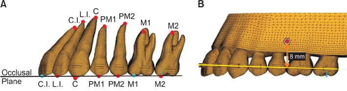
Figure 1 Three-dimensional finite element model. A, Landmarks for the assessment of displacement; B, location of the miniscrew. Dots indicate points for (mesiobuccal) cusp tips/middle incisal edge or mesial root apices. Miniscrew position was simulated at 8 mm above the main archwire, at the contact point portion between the 2nd premolar and 1st molar. C.I., central incisor; L.I., lateral incisor; C, canine; PM1, 1st premolar; PM2, 2nd premolar; M1, 1st molar; M2, 2nd molar.
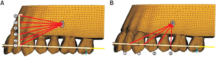
Figure 2 Experimental conditions. A, Conditions 1-6: effect of lever arm length (extended between lateral incisor and canine, conditions 1: 0 mm, 2: 2 mm, 3: 4 mm, 4: 6 mm, 5: 8 mm, 6: 10 mm, respectively). B, Conditions 1, and 7-9: effect of force application point (conditions 7: between canine and 1st premolar, 8: between 1st and 2nd premolars, 9: between 2nd premolar and 1st molar).
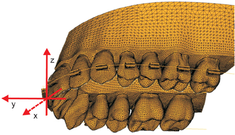
Figure 3 Coordinate system. x-axis: (+) palatal, (-) buccal direction; y-axis: (+) anterior, (-) posterior direction; z-axis: (+) superior, (-) inferior direction.
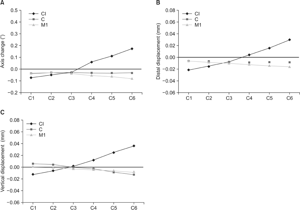
Figure 4 Effect of lever arm length in conditions 1-6 (C1-6). A, Axis change of the central incisor (CI), canine (C), and first molar (M1). B, Distal displacement of CI, C, and M1. C, Vertical displacement of CI, C, and M1. C1-6: conditions 1-6 (for definition, refer to Figure 2).
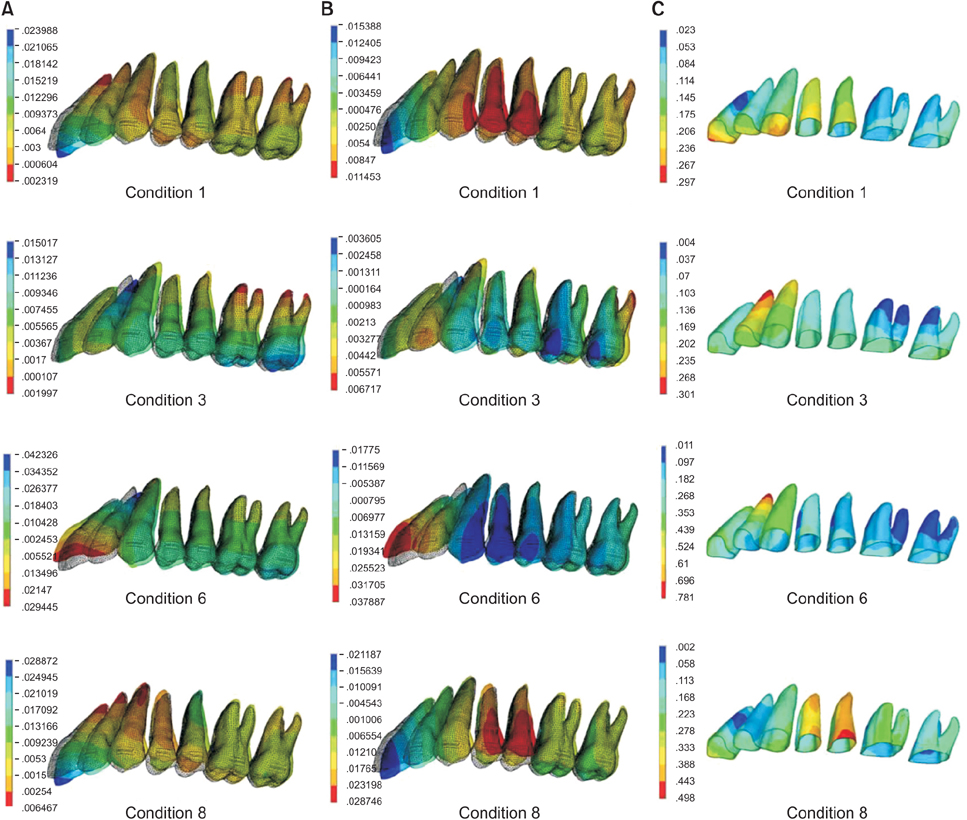
Figure 5 Contour plots. A, Distal displacement (mm). B, Vertical displacement (mm). C, von Mises stress distribution (g/mm2) in conditions 1, 3, 6, and 8.
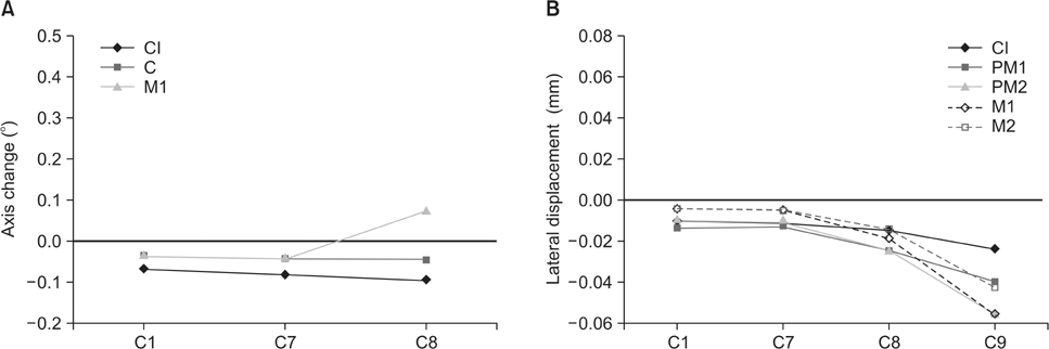
Figure 6 Effect of force application point in conditions 1, and 7-9 (C1, C7-9). A, Axis change of the central incisor (CI), canine (C), and first molar (M1). B, Lateral displacement of the CI, first premolar (PM1), second premolar (PM2), M1, and second molar (M2). C1, 7 and 8: conditions 1, 7 and 8 (for the definition, refer to Figure 2).
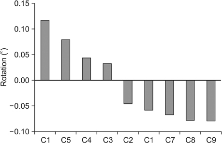
Figure 7 Rotation of the occlusal plane in conditions 1-9, presented in descending order of the degree of rotation. (+): flattening rotation; (-) steepening rotation. For definition of C1 to C9, refer to Figure 2.
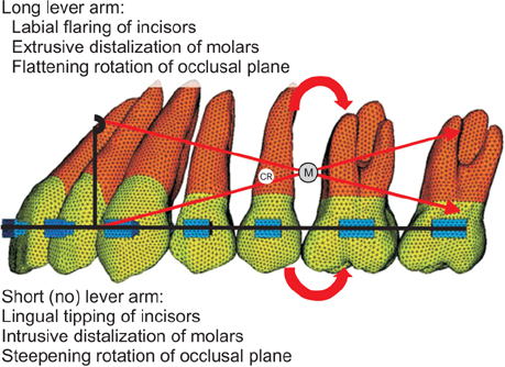
Figure 8 Schematic guidelines for the distalization of the entire maxillary arch. M, miniscrew; CR, center of the resistance of the whole maxillary dentition estimated by Jeong et al.23
Cited by 9 articles
-
Effect of labiolingual inclination of a maxillary central incisor and surrounding alveolar bone loss on periodontal stress: A finite element analysis
Sung-Hwan Choi, Young-Hoon Kim, Kee-Joon Lee, Chung-Ju Hwang
Korean J Orthod. 2016;46(3):155-162. doi: 10.4041/kjod.2016.46.3.155.Finite element analysis of maxillary incisor displacement during en-masse retraction according to orthodontic mini-implant position
Jae-Won Song, Joong-Ki Lim, Kee-Joon Lee, Sang-Jin Sung, Youn-Sic Chun, Sung-Seo Mo
Korean J Orthod. 2016;46(4):242-252. doi: 10.4041/kjod.2016.46.4.242.The effects of alveolar bone loss and miniscrew position on initial tooth displacement during intrusion of the maxillary anterior teeth: Finite element analysis
Sun-Mi Cho, Sung-Hwan Choi, Sang-Jin Sung, Hyung-Seog Yu, Chung-Ju Hwang
Korean J Orthod. 2016;46(5):310-322. doi: 10.4041/kjod.2016.46.5.310.Three-dimensional evaluation of tooth movement in Class II malocclusions treated without extraction by orthodontic mini-implant anchorage
Dler Ali, Hnd Mohammed, Seung-Hwan Koo, Kyung-Hwa Kang, Sang-Cheol Kim
Korean J Orthod. 2016;46(5):280-289. doi: 10.4041/kjod.2016.46.5.280.A three-dimensional finite element analysis of molar distalization with a palatal plate, pendulum, and headgear according to molar eruption stage
Ju-Man Kang, Jae Hyun Park, Mohamed Bayome, Moonbee Oh, Chong Ook Park, Yoon-Ah Kook, Sung-Seo Mo
Korean J Orthod. 2016;46(5):290-300. doi: 10.4041/kjod.2016.46.5.290.Finite-element analysis of the center of resistance of the mandibular dentition
A-Ra Jo, Sung-Seo Mo, Kee-Joon Lee, Sang-Jin Sung, Youn-Sic Chun
Korean J Orthod. 2017;47(1):21-30. doi: 10.4041/kjod.2017.47.1.21.Biomechanical analysis of distalization of mandibular molars by placing a mini-plate: A finite element study
Myungsoon Park, Yonghyun Na, Minbong Park, Janghoon Ahn
Korean J Orthod. 2017;47(5):289-297. doi: 10.4041/kjod.2017.47.5.289.Type of tooth movement during en masse retraction of the maxillary anterior teeth using labial versus lingual biocreative therapy in adults: A randomized clinical trial
Mais M. Sadek, Noha E. Sabet, Islam T. Hassan
Korean J Orthod. 2019;49(6):381-392. doi: 10.4041/kjod.2019.49.6.381.Total intrusion and distalization of the maxillary arch to improve smile esthetics
Eui Seon Baek, Soonshin Hwang, Kyung-Ho Kim, Chooryung J. Chung
Korean J Orthod. 2017;47(1):59-73. doi: 10.4041/kjod.2017.47.1.59.
Reference
-
1. Lee KJ, Park YC, Hwang WS, Seong EH. Uprighting mandibular second molars with direct miniscrew anchorage. J Clin Orthod. 2007; 41:627–635.2. Park YC, Lee SY, Kim DH, Jee SH. Intrusion of posterior teeth using mini-screw implants. Am J Orthod Dentofacial Orthop. 2003; 123:690–694.
Article3. Yamada K, Kuroda S, Deguchi T, Takano-Yamamoto T, Yamashiro T. Distal movement of maxillary molars using miniscrew anchorage in the buccal interradicular region. Angle Orthod. 2009; 79:78–84.
Article4. Byloff FK, Darendeliler MA. Distal molar movement using the pendulum appliance. Part 1: Clinical and radiological evaluation. Angle Orthod. 1997; 67:249–260.5. Tominaga JY, Tanaka M, Koga Y, Gonzales C, Kobayashi M, Yoshida N. Optimal loading conditions for controlled movement of anterior teeth in sliding mechanics. Angle Orthod. 2009; 79:1102–1107.
Article6. Lee KJ, Park YC, Hwang CJ, Kim YJ, Choi TH, Yoo HM, et al. Displacement pattern of the maxillary arch depending on miniscrew position in sliding mechanics. Am J Orthod Dentofacial Orthop. 2011; 140:224–232.
Article7. Chaconas SJ, Caupto AA, Miyashita K. Force distribution comparisons of various retraction archwires. Angle Orthod. 1989; 59:25–30.8. Billiet T, de Pauw G, Dermaut L. Location of the centre of resistance of the upper dentition and the nasomaxillary complex. An experimental study. Eur J Orthod. 2001; 23:263–273.
Article9. Pedersen E, Andersen K, Melsen B. Tooth displacement analysed on human autopsy material by means of a strain gauge technique. Eur J Orthod. 1991; 13:65–74.
Article10. Tanne K, Nagataki T, Inoue Y, Sakuda M, Burstone CJ. Patterns of initial tooth displacements associated with various root lengths and alveolar bone heights. Am J Orthod Dentofacial Orthop. 1991; 100:66–71.
Article11. Mo SS, Kim SH, Sung SJ, Chung KR, Chun YS, Kook YA, et al. Factors controlling anterior torque with C-implants depend on en-masse retraction without posterior appliances: biocreative therapy type II technique. Am J Orthod Dentofacial Orthop. 2011; 139:e183–e191.
Article12. Andrews LF. The six keys to normal occlusion. Am J Orthod. 1972; 62:296–309.
Article13. Coolidge E. The thickness of the human periodontal membrane. J Am Dent Assoc. 1937; 24:1260–1267.
Article14. Block PL. Restorative margins and periodontal health: a new look at an old perspective. J Prosthet Dent. 1987; 57:683–689.
Article15. Sung SJ, Baik HS, Moon YS, Yu HS, Cho YS. A comparative evaluation of different compensating curves in the lingual and labial techniques using 3D FEM. Am J Orthod Dentofacial Orthop. 2003; 123:441–450.
Article16. Reimann S, Keilig L, Jäger A, Bourauel C. Biomechanical finite-element investigation of the position of the centre of resistance of the upper incisors. Eur J Orthod. 2007; 29:219–224.
Article17. Tanne K, Sakuda M, Burstone CJ. Three-dimensional finite element analysis for stress in the periodontal tissue by orthodontic forces. Am J Orthod Dentofacial Orthop. 1987; 92:499–505.
Article18. Burstone CJ, Koenig HA. Force systems from an ideal arch. Am J Orthod. 1974; 65:270–289.
Article19. Cattaneo PM, Dalstra M, Melsen B. The finite element method: a tool to study orthodontic tooth movement. J Dent Res. 2005; 84:428–433.
Article20. Gröning F, Fagan MJ, O'Higgins P. The effects of the periodontal ligament on mandibular stiffness: a study combining finite element analysis and geometric morphometrics. J Biomech. 2011; 44:1304–1312.
Article21. McGuinness NJ, Wilson AN, Jones ML, Middleton J. A stress analysis of the periodontal ligament under various orthodontic loadings. Eur J Orthod. 1991; 13:231–242.
Article22. Andersen KL, Pedersen EH, Melsen B. Material parameters and stress profiles within the periodontal ligament. Am J Orthod Dentofacial Orthop. 1991; 99:427–440.
Article23. Jeong GM, Sung SJ, Lee KJ, Chun YS, Mo SS. Finite-element investigation of the center of resistance of the maxillary dentition. Korean J Orthod. 2009; 39:83–94.
Article24. Volchansky A, Cleaton-Jones P. Clinical crown height (length)--a review of published measurements. J Clin Periodontol. 2001; 28:1085–1090.
Article25. Tenenbaum H, Tenenbaum M. A clinical study of the width of the attached gingiva in the deciduous, transitional and permanent dentitions. J Clin Periodontol. 1986; 13:270–275.
Article26. Bechtold TE, Kim JW, Choi TH, Park YC, Lee KJ. Distalization pattern of the maxillary arch depending on the number of orthodontic miniscrews. Angle Orthod. 2013; 83:266–273.
Article27. Choy K, Pae EK, Park Y, Kim KH, Burstone CJ. Effect of root and bone morphology on the stress distribution in the periodontal ligament. Am J Orthod Dentofacial Orthop. 2000; 117:98–105.
Article28. Baek MS, Choi YJ, Yu HS, Lee KJ, Kwak J, Park YC. Long-term stability of anterior open-bite treatment by intrusion of maxillary posterior teeth. Am J Orthod Dentofacial Orthop. 2010; 138:396.e1–396.e9. discussion 396-8.
Article29. Park HS, Kwon OW, Sung JH. Nonextraction treatment of an open bite with microscrew implant anchorage. Am J Orthod Dentofacial Orthop. 2006; 130:391–402.
Article30. Park HS, Kwon TG, Sung JH. Nonextraction treatment with microscrew implants. Angle Orthod. 2004; 74:539–549.
- Full Text Links
-
- Actions
-
Cited
- CITED
-
- Close
- Share
-
- Similar articles
-
- En masse maxillary dentition distalization with clear aligners using infrazygomatic mini-implants: A finite element study
- Immediate changes in the mandibular dentition after maxillary molar distalization using headgear
- Biomechanical analysis for different mandibular total distalization methods with clear aligners: A finite element study
- Total arch distalization with interproximal stripping in a patient with severe crowding
- Total intrusion and distalization of the maxillary arch to improve smile esthetics
