Korean J Orthod.
2017 Mar;47(2):142-147. 10.4041/kjod.2017.47.2.142.
Immediate changes in the mandibular dentition after maxillary molar distalization using headgear
- Affiliations
-
- 1Department of Orthodontics, School of Dentistry, Chonnam National University, Gwangju, Korea. ortholkm@chonnam.ac.kr
- KMID: 2400391
- DOI: http://doi.org/10.4041/kjod.2017.47.2.142
Abstract
- The purpose of this study was to investigate immediate changes in the mandibular dentition after maxillary molar distalization using headgear in non-growing patients. Sixteen patients (mean age, 18.9 ± 2.0 years) with Class II molar relationship and crowding were included in the present study. To correct the molar relationship, headgear was used for maxillary molar distalization. Cone-beam computed tomography-generated half-cephalograms (CG Cephs) and dental casts were used to evaluate dental changes for each subject before and immediately after molar distalization using headgear. The mean duration that subjects wore the headgear was 6.3 months. CG Cephs showed that the first maxillary molars were distalized 4.2 ± 1.6 mm with 9.7°± 6.1° of distal angulation. The intercanine, interpremolar, and intermolar widths of the mandible increased after maxillary molar distalization. The present study's results suggest that maxillary molar distalization using headgear induces a spontaneous response in the untreated mandibular dentition of non-growing patients.
Keyword
MeSH Terms
Figure
-
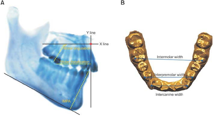
Figure 1 Measurements used in this study. A, Cone-beam computed tomography-generated half-cephalogram (CG Ceph) measurements for maxillary molar distalization, maxillary molar angulation, and mandibular incisor inclination (IMPA). On the CG Ceph, a horizontal reference line (X line) was established parallel to the head posture aligner after 3-dimensional superimposition of the two volume data sets. The vertical reference line (Y line) was perpendicular to the horizontal reference line (X line), which passed through A point. B, Model measurements for assessing the mandibular dental changes.
Reference
-
1. Bae SM, Park HS, Kyung HM, Kwon OW, Sung JH. Clinical application of micro-implant anchorage. J Clin Orthod. 2002; 36:298–302.2. Nishimura M, Sannohe M, Nagasaka H, Igarashi K, Sugawara J. Nonextraction treatment with temporary skeletal anchorage devices to correct a Class II Division 2 malocclusion with excessive gingival display. Am J Orthod Dentofacial Orthop. 2014; 145:85–94.
Article3. Kook YA, Bayome M, Trang VT, Kim HJ, Park JH, Kim KB, et al. Treatment effects of a modified palatal anchorage plate for distalization evaluated with cone-beam computed tomography. Am J Orthod Dentofacial Orthop. 2014; 146:47–54.
Article4. Kuroda S, Yamada K, Deguchi T, Hashimoto T, Kyung HM, Takano-Yamamoto T. Root proximity is a major factor for screw failure in orthodontic anchorage. Am J Orthod Dentofacial Orthop. 2007; 131:4 Suppl. S68–S73.
Article5. Miyazawa K, Kawaguchi M, Tabuchi M, Goto S. Accurate pre-surgical determination for self-drilling miniscrew implant placement using surgical guides and cone-beam computed tomography. Eur J Orthod. 2010; 32:735–740.
Article6. Feldmann I, List T, Feldmann H, Bondemark L. Pain intensity and discomfort following surgical placement of orthodontic anchoring units and premolar extraction: a randomized controlled trial. Angle Orthod. 2007; 77:578–585.
Article7. Garfinkle JS, Cunningham LL Jr, Beeman CS, Kluemper GT, Hicks EP, Kim MO. Evaluation of orthodontic mini-implant anchorage in premolar extraction therapy in adolescents. Am J Orthod Dentofacial Orthop. 2008; 133:642–653.
Article8. Cangialosi TJ, Meistrell ME Jr, Leung MA, Ko JY. A cephalometric appraisal of edgewise Class II nonextraction treatment with extraoral force. Am J Orthod Dentofacial Orthop. 1988; 93:315–324.
Article9. Mossaz CF, Byloff FK, Kiliaridis S. Cervical headgear vs pendulum appliance for the treatment of moderate skeletal Class II malocclusion. Am J Orthod Dentofacial Orthop. 2007; 132:616–623.
Article10. Freitas MR, Lima DV, Freitas KM, Janson G, Henriques JF. Cephalometric evaluation of Class II malocclusion treatment with cervical headgear and mandibular fixed appliances. Eur J Orthod. 2008; 30:477–482.
Article11. Sfondrini MF, Cacciafesta V, Sfondrini G. Upper molar distalization: a critical analysis. Orthod Craniofac Res. 2002; 5:114–126.
Article12. Proffit WP, Fields HW, Sarver DM. Orthodontic treatment planning: from problem list to specific plan. In : Proffit WP, Fields HW, Sarver DM, editors. Contemporary orthodontics. 5th ed. St. Louis: Elsevier-Mosby;2013. p. 220–277.13. Proffit WP. Retention. In : Proffit WP, Fields HW, Sarver DM, editors. Contemporary orthodontics. 5th ed. St. Louis: Elsevier-Mosby;2013. p. 622–622. .14. Fields HW, Proffit WP. Treatment of skeletal problems in children and preadolescents. In : Proffit WP, Fields HW, Sarver DM, editors. Contemporary orthodontics. 5th ed. St. Louis: Elsevier-Mosby;2013. p. 472–528.15. Hwang HS, Lee KM, Uhm GS, Cho JH, McNamara JA Jr. Use of Reference Ear Plug to improve accuracy of lateral cephalograms generated from cone-beam computed tomography scans. Korean J Orthod. 2013; 43:54–61.
Article16. Lee KM, Lee WJ, Cho JH, Hwang HS. Three-dimensional prediction of the nose for facial reconstruction using cone-beam computed tomography. Forensic Sci Int. 2014; 236:194.e1–194.e5.
Article17. Cha BK, Ngan PW. Skeletal anchorage for orthopedic correction of growing class III patients. Semin Orthod. 2011; 17:124–137.
Article18. Faul F, Erdfelder E, Lang AG, Buchner A. G*Power 3: a flexible statistical power analysis program for the social, behavioral, and biomedical sciences. Behav Res Methods. 2007; 39:175–191.
Article19. Dahlberg G. Statistical methods for medical and biological students. London: George Allen & Unwin Ltd;1940. p. 122–132.20. Kirjavainen M, Kirjavainen T, Haavikko K. Changes in dental arch dimensions by use of an orthopedic cervical headgear in Class II correction. Am J Orthod Dentofacial Orthop. 1997; 111:59–66.
Article21. Proffit WP. The etiology of orthodontic problems. In : Proffit WP, Fields HW, Sarver DM, editors. Contemporary orthodontics. 5th ed. St. Louis: Elsevier-Mosby;2013. p. 114–149.22. Miller CL, Araújo EA, Behrents RG, Oliver DG, Tanaka OM. Mandibular arch dimensions following bonded and banded rapid maxillary expansion. J World Fed Orthod. 2014; 3:119–123.
Article
- Full Text Links
-
- Actions
-
Cited
- CITED
-
- Close
- Share
-
- Similar articles
-
- Combined treatment with headgear and the Frog appliance for maxillary molar distalization: a randomized controlled trial
- The effects of high pull headgear in mixed dention with Class II malocclusion
- A cephalometric evaluation of anterior J hook headgear traction to the maxilla
- A three-dimensional finite element analysis of molar distalization with a palatal plate, pendulum, and headgear according to molar eruption stage
- Treatment effects of the Teuscher appliance in skeletal Class II division 1 malocclusion
