Korean J Physiol Pharmacol.
2009 Jun;13(3):251-256. 10.4196/kjpp.2009.13.3.251.
Bis Is Involved in Glial Differentiation of P19 Cells Induced by Retinoic Acid
- Affiliations
-
- 1Department of Biomedical Science, Graduate School, College of Medicine, The Catholic University of Korea, Seoul 137-701, Korea.
- 2Department of Anatomy, Graduate School, College of Medicine, The Catholic University of Korea, Seoul 137-701, Korea.
- 3Division of Radiation Cancer Research, Korea Institute of Radiological and Medical Sciences, Seoul 139-706, Korea.
- 4Bioindustry Research Center, Korea Research Institute Bioscience and Biotechnology, Daejeon 305-806, Korea.
- 5Department of Internal Medicine, College of Medicine, The Catholic University of Korea, Seoul 137-701, Korea. leejh@catholic.ac.kr
- 6Department of Biochemistry, College of Medicine, The Catholic University of Korea, Seoul 137-701, Korea. leejh@catholic.ac.kr
- KMID: 2071678
- DOI: http://doi.org/10.4196/kjpp.2009.13.3.251
Abstract
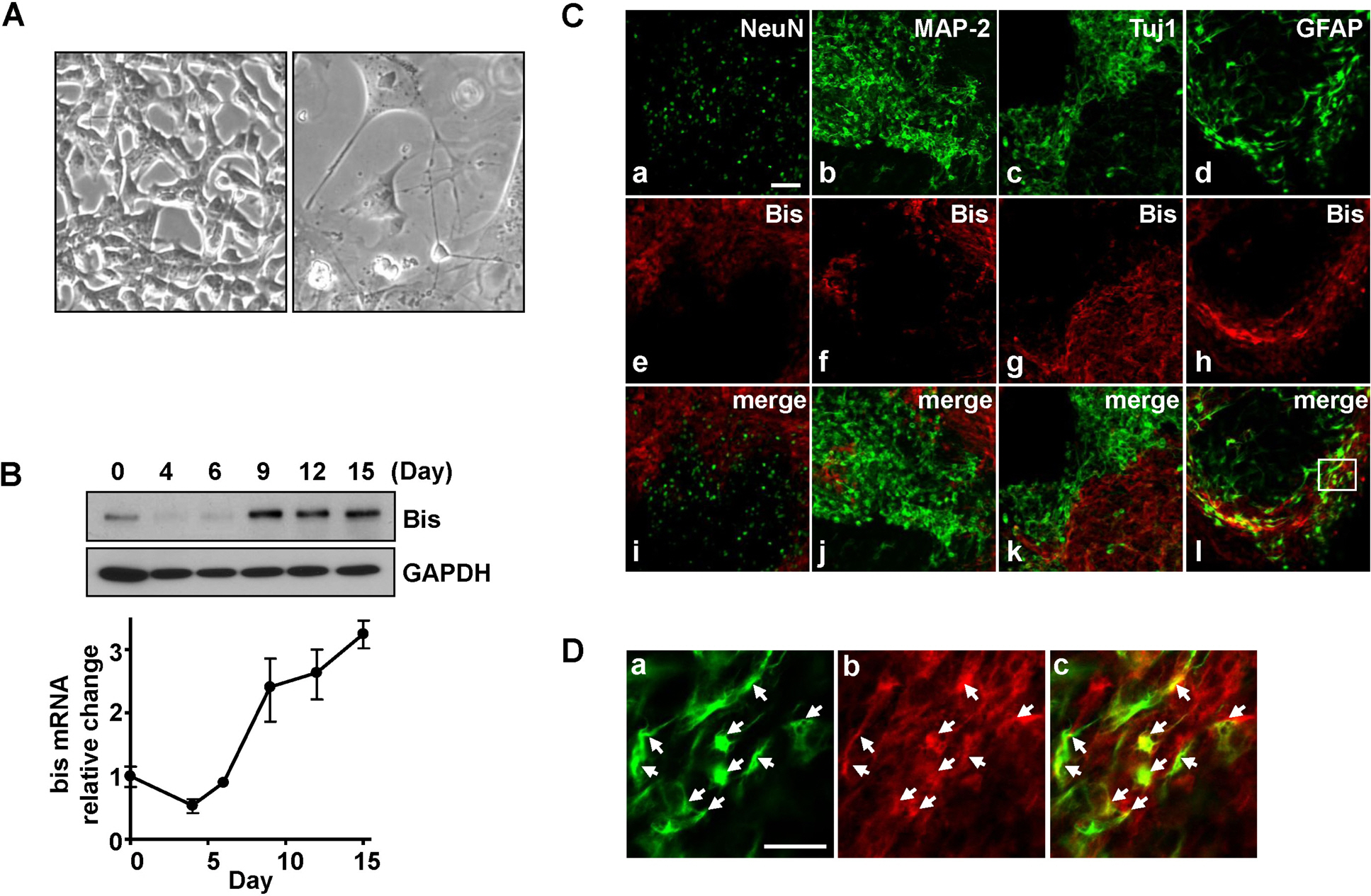
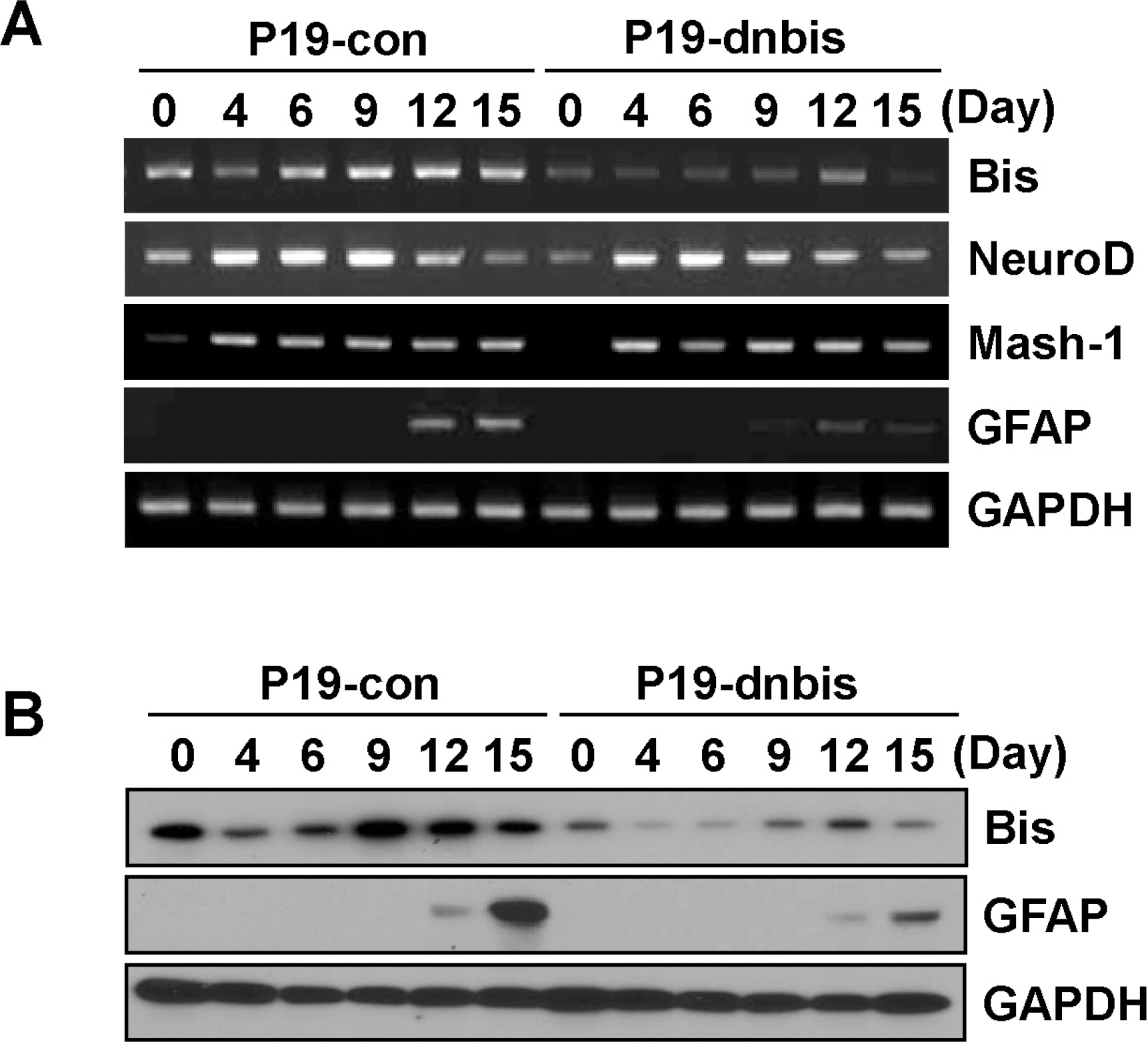
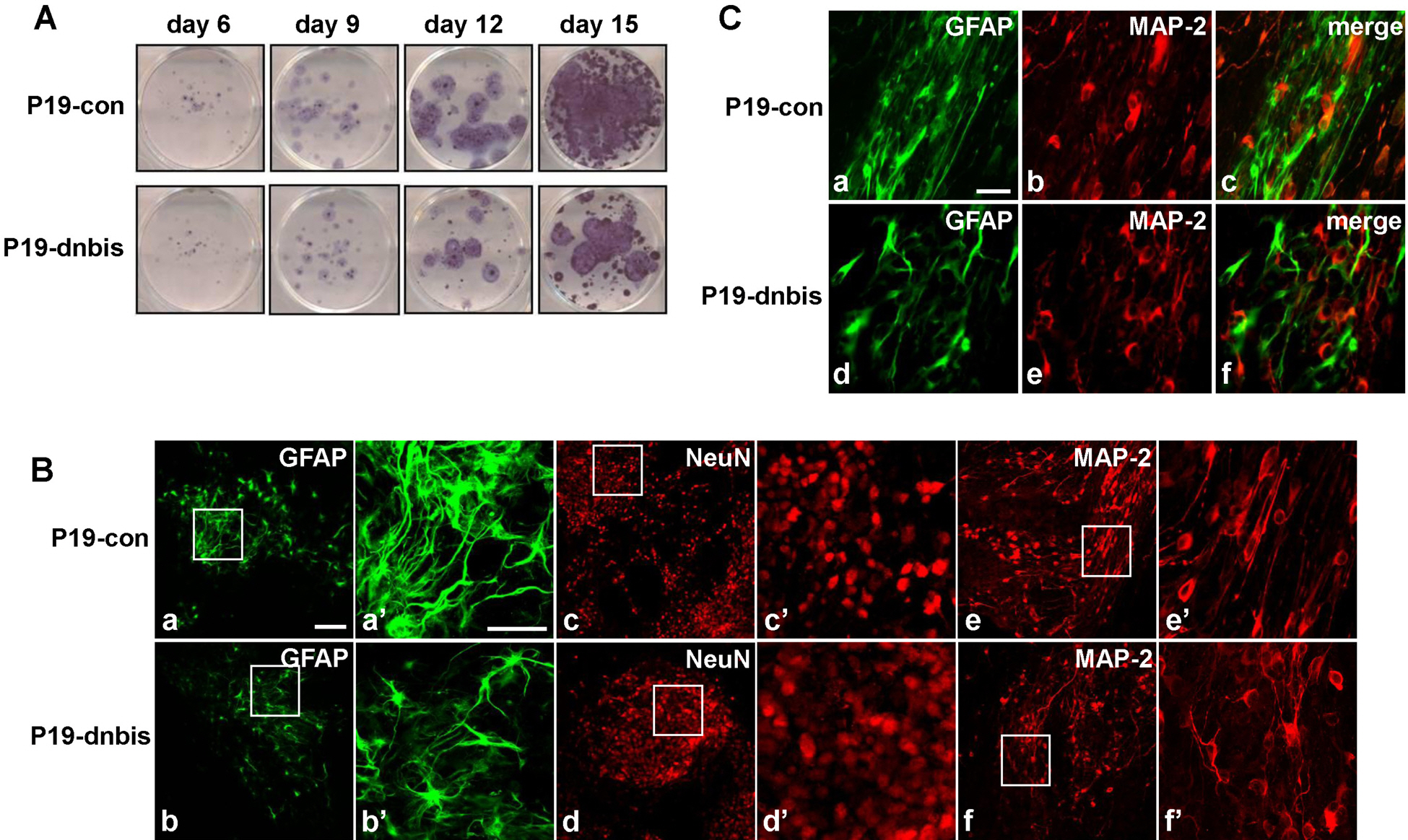
- Previous observations suggest that Bis, a Bcl-2-binding protein, may play a role the neuronal and glial differentiation in vivo. To examine this further, we investigated Bis expression during the in vitro differentiation of P19 embryonic carcinoma cells induced by retinoic acid (RA). Western blotting and RT-PCR assays showed that Bis expression was temporarily decreased during the free floating stage and then began to increase on day 6 after the induction of differentiation. Double immunostaining indicated that Bis-expressing cells do not express several markers of differentiation, including NeuN, MAP-2 and Tuj-1. However, some of the Bis-expressing cells also were stained with GFAP-antibodies, indicating that Bis is involved glial differentiation. Using an shRNA strategy, we developed bis-knock down P19 cells and compared them with control P19 cells for the expression of NeuroD, Mash-1 and GFAP during RA-induced differentiation. Among these, only GFAP induction was significantly attenuated in P19-dnbis cells and the population showing GFAP immunoreactivity was also decreased. It is noteworthy that distribution of mature neurons and migrating neurons was disorganized, and the close association of migrating neuroblasts with astrocytes was not observed in P19-dnbis cells. These results suggest that Bis is involved in the migration-inducing activity of glial cells.
Keyword
MeSH Terms
Figure
Cited by 2 articles
-
The Effect of Metformin Treatment on CRBP-I Level and Cancer Development in the Liver of HBx Transgenic Mice
Jo-Heon Kim, Md. Morshedul Alam, Doek Bae Park, Moonjae Cho, Seung-Hong Lee, You-Jin Jeon, Dae-Yeul Yu, Tae Du Kim, Ha Young Kim, Chung Gu Cho, Dae Ho Lee
Korean J Physiol Pharmacol. 2013;17(5):455-461. doi: 10.4196/kjpp.2013.17.5.455.Bis is Induced by Oxidative Stress
via Activation of HSF1
Hyung Jae Yoo, Chang-Nim Im, Dong-Ye Youn, Hye Hyeon Yun, Jeong-Hwa Lee
Korean J Physiol Pharmacol. 2014;18(5):403-409. doi: 10.4196/kjpp.2014.18.5.403.
Reference
-
Bonelli P., Petrella A., Rosati A., Romano MF., Lerose R., Pagliuca MG., Amelio T., Festa M., Martire G., Venuta S., Turco MC., Leone A. BAG3 protein regulates stress-induced apoptosis in normal and neoplastic leukocytes. Leukemia. 18:358–360. 2004.
ArticleCarra S., Seguin SJ., Lambert H., Landry J. HspB8 chaperone activity toward poly(Q)-containing proteins depends on its association with Bag3, a stimulator of macroautophagy. J Biol Chem. 283:1437–1444. 2008.
ArticleChen L., Wu W., Dentchev T., Zeng Y., Wang J., Tsui I., Tobias JW., Bennett J., Baldwin D., Dunaief JL. Light damage induced changes in mouse retinal gene expression. Exp Eye Res. 79:239–247. 2004.
ArticleChoi JS., Lee JH., Kim HY., Chun MH., Chung JW., Lee MY. Developmental expression of Bis protein in the cerebral cortex and hippocampus of rats. Brain Res. 1092:69–78. 2006.
ArticleChoi JS., Lee JH., Shin YJ., Lee JY., Yun H., Chun MH., Lee MY. Transient expression of Bis protein in the midline glial structure in the developing rat brainstem and spinal cord. Cell Tissue Res. 337:27–36. 2009.Doong H., Price J., Kim YS., Gasbarre C., Probst J., Liotta LA., Blanchette J., Rizzo K., Kohn E. CAIR-1/BAG-3 forms an EGF-regulated ternary complex with phospholipase C-gamma and Hsp70/Hsc70. Oncogene. 19:4385–4395. 2000.Hatten ME. Central nervous system neuronal migration. Annu Rev Neurosci. 22:511–539. 1999.
ArticleHong S., Heo J., Lee S., Heo S., Kim SS., Lee YD., Kwon M. Methyltransferase-inhibition interferes with neuronal differentiation of P19 embryonal carcinoma cells. Biochem Biophys Res Commun. 377:935–940. 2008.
ArticleIwasaki M., Homma S., Hishiya A., Dolezal SJ., Reed JC., Takayama S. BAG3 regulates motility and adhesion of epithelial cancer cells. Cancer Res. 67:10252–10259. 2007.
ArticleJacobs AT., Marnett LJ. HSF1-mediated BAG3 expression attenuates apoptosis in 4-hydroxynonenal-treated colon cancer cells via stabilization of anti-apoptotic Bcl-2 proteins. J Biol Chem. 284:9176–9183. 2009.
ArticleJing XT., Wu HT., Wu Y., Ma X., Liu SH., Wu YR., Ding XF., Peng XZ., Qiang BQ., Yuan JG., Fan WH., Fan M. DIXDC1 promotes retinoic acid-induced neuronal differentiation and inhibits gliogenesis in P19 cells. Cell Mol Neurobiol. 29:55–67. 2009.
ArticleJones-Villeneuve EM., McBurney MW., Rogers KA., Kalnins VI. Retinoic acid induces embryonal carcinoma cells to differentiate into neurons and glial cells. J Cell Biol. 94:253–262. 1982.
ArticleJones-Villeneuve EM., Rudnicki MA., Harris JF., McBurney MW. Retinoic acid-induced neural differentiation of embryonal carcinoma cells. Mol Cell Biol. 3:2271–2279. 1983.
ArticleKassis JN., Guancial EA., Doong H., Virador V., Kohn EC. CAIR-1/BAG-3 modulates cell adhesion and migration by downregulating activity of focal adhesion proteins. Exp Cell Res. 312:2962–2971. 2006.
ArticleLee JH., Takahashi T., Yasuhara N., Inazawa J., Kamada S., Tsujimoto Y. Bis, a Bcl-2-binding protein that synergizes with Bcl-2 in preventing cell death. Oncogene. 18:6183–6190. 1999.
ArticleLee MY., Kim SY., Shin SL., Choi YS., Lee JH., Tsujimoto Y. Reactive astrocytes express bis, a bcl-2-binding protein, after transient forebrain ischemia. Exp Neurol. 175:338–346. 2002.
ArticleLim DA., Alvarez-Buylla A. Interaction between astrocytes and adult subventricular zone precursors stimulates neurogenesis. Proc Natl Acad Sci U S A. 96:7526–7531. 1999.
ArticleLiour SS., Yu RK. Differentiation of radial glia-like cells from embryonic stem cells. Glia. 42:109–117. 2003.
ArticlePagliuca MG., Lerose R., Cigliano S., Leone A. Regulation by heavy metals and temperature of the human BAG-3 gene, a modulator of Hsp70 activity. FEBS Lett. 541:11–15. 2003.
ArticlePark HJ., Choi JS., Chun MH., Chung JW., Jeon MH., Lee JH., Lee MY. Immunohistochemical localization of Bis protein in the rat central nervous system. Cell Tissue Res. 314:207–214. 2003.
ArticleRomano MF., Festa M., Pagliuca G., Lerose R., Bisogni R., Chiurazzi F., Storti G., Volpe S., Venuta S., Turco MC., Leone A. BAG3 protein controls B-chronic lymphocytic leukaemia cell apoptosis. Cell Death Differ. 10:383–385. 2003.
ArticleSantiago MF., Liour SS., Mendez-Otero R., Yu RK. Glial-guided neuronal migration in P19 embryonal carcinoma stem cell aggregates. J Neurosci Res. 81:9–20. 2005.
ArticleTakayama S., Xie Z., Reed JC. An evolutionarily conserved family of Hsp70/Hsc70 molecular chaperone regulators. J Biol Chem. 274:781–786. 1999.
Article
- Full Text Links
- Actions
-
Cited
- CITED
-
- Close
- Share
- Similar articles
-
- Neuronal Differentiatin of P19 Cells
- Induction of Embryonal Teratocarcinoma Cell(P19 cell) to Differentiate into Smooth Muscle Cell
- NgR1 Expressed in P19 Embryonal Carcinoma Cells Differentiated by Retinoic Acid Can Activate STAT3
- The Effect of Intravitreal Injection of Retinoic Acid on Experimentally Induced Proliferative Vitreoretinopathy using Human Retinal Prgment Epithelium in Rabbits
- Studies on Functional Differentiation of Small Intestinal Epithelial Cells


